Impact Factor ISSN: 1837-9664




J Cancer 2026; 17(11):1965-1977. doi:10.7150/jca.144312 This issue Cite
Review
Who Should Undergo Surgery, and at What Operative Risk? Two Conditions Governing Cytoreduction in TKI-Treated Gastrointestinal Stromal Tumor
Chun-Nan Yeh, MD 
GIST Team, Department of Surgery, Chang Gung Memorial Hospital, Linkou; Chang Gung University, Taoyuan, Taiwan.
Received 2026-9-17; Accepted 2026-10-15; Published 2026-10-31
Abstract
Gastrointestinal stromal tumor is where targeted therapy was first shown to work, and where the surgical question remains least resolved. Two randomized trials of cytoreductive surgery closed on accrual for a structural reason: both required randomization at best radiological response, the moment at which individual equipoise collapses. The literature that filled the gap is uniformly selected and is commonly set aside on that ground.
Setting it aside overlooks a distinction that matters clinically. Response to tyrosine kinase inhibitor (TKI) therapy determines both who is offered an operation and how long a patient survives, so it confounds every observational estimate; it is also a clinical read-out of clonal state, and a plausible modifier of surgical effect. The task is not to adjust response away but to condition the question on it.
Two conditions should jointly govern the decision. The first is biological: whether surgery can return the tumor to systemic control, which turns on the proportion of tumor burden that has escaped drug control rather than on clone number. The second is operative morbidity: preoperative TKI exposure impairs tissue repair and raises morbidity and mortality, and complications cost time on systemic therapy, itself a treatment failure in a disease controlled by continuous kinase inhibition. Focal progression is best supported; consolidating a complete response is plausible but unproven; prophylactic resection is a hypothesis; generalized progression shows no benefit. The framework is hypothesis-generating, and four questions retaining equipoise are specified, of which randomizing the perioperative interruption interval is most deliverable.
Keywords: gastrointestinal stromal tumor, cytoreductive surgery, tyrosine kinase inhibitor, secondary KIT mutation, clonal resistance, treatment interruption
1. Introduction
Within three years of the identification of activating KIT mutations as the driver of gastrointestinal stromal tumor (GIST), a kinase inhibitor had converted a chemoresistant sarcoma with a median survival of approximately eighteen months into a disease routinely controlled for years [1-4]. The surgical consequences have never been settled. Before 2001 surgery was the treatment of GIST wherever feasible; after 2001 it became one component of a drug-defined strategy, an adjunct whose indication, timing and extent depend on how the tumor is behaving under systemic therapy. The evidence base did not change: it remains retrospective and, at its largest, an analysis of 323 patients undergoing 400 operations at two centers [5]; the largest single-institution surgical experience, 1000 patients spanning the pre- and post-imatinib eras, is similarly retrospective [6].
The standard criticism is that these cohorts are chosen: patients offered cytoreduction are those responding, those fit for laparotomy, and those whose disease is anatomically approachable. The criticism is correct as far as it goes, and it has kept surgery out of guideline recommendations for advanced disease in most jurisdictions [7-10].
It is nonetheless incomplete, and this review advances three propositions. First, response to TKI therapy is simultaneously a source of confounding and a biologically meaningful effect modifier: it predicts who is offered an operation and how long they live, and appears to be a clinical read-out of clonal state. Treating it only as a nuisance variable discards the information it carries; treating it only as an indication ignores the confounding it produces. Second, the benefit of an operation cannot be assessed separately from the morbidity prior systemic therapy imposes, and that morbidity has an oncological price paid in interrupted drug exposure. Third, the indication should therefore require two conditions rather than one.
Because these propositions rest on evidence of unequal strength, three levels are distinguished throughout: level A, an observed clinical association; level B, mechanistic support; level C, causal evidence from randomization, which for the central question does not exist. Levels A and B suffice to formulate a decision hypothesis but do not substitute for level C. The framework applies to elective cytoreduction in advanced, KIT- or PDGFRA-driven GIST under TKI therapy. Operations undertaken for hemorrhage, perforation or obstruction are excluded throughout: the indication there is the emergency, no selection decision exists, and life-saving intervention takes precedence over every consideration below. SDH-deficient and other TKI-insensitive tumors follow a different logic and are treated as an explicit exception.
2. Why the randomized question failed
Two randomized trials have addressed whether cytoreduction adds anything to continued TKI therapy in responding metastatic disease. EORTC 62063 was designed for 350 patients across sixty centers and closed for want of accrual. A Chinese multicenter trial closed early with 41 patients randomized, reporting two-year progression-free survival of 88.4% versus 57.7% (P = 0.089) and median overall survival not reached versus 49 months (P = 0.024) [11].
The conventional reading is that the question is unanswered because the trials were unlucky. Both protocols required randomization at or near best response, timing forced by the biology, since operating before the drug has selected the responsive disease removes lesions that would have melted and leaves those that would not [12, 13]. But best response is also the moment at which uncertainty resolves for the two people who must consent: community equipoise persists, individual equipoise does not. This is a structural property of any disease in which a drug produces deep, durable responses in most patients, and it will not be solved with more centers. It changes what a review can usefully do — not await the definitive trial, but establish what the non-randomizable evidence can and cannot support.
3. The first condition: biological opportunity
3.1 The response hierarchy and the meaning of its reproducibility
Every series stratifying cytoreduction by disease status at operation has recovered the same ordering: partial response or stable disease does best, focal progression nearly as well, generalized progression not at all [5, 14-17]. In the largest dataset, the imatinib subset gave median progression-free survival from operation of 36, 30, 11 and 6 months for responsive, stable, unifocal-progressive and multifocal-progressive disease, with median overall survival not reached, 110, 59 and 24 months; multifocal progression was independently prognostic of worse progression-free (hazard ratio 3.08) and overall survival (hazard ratio 5.84) [5].
Grouping partial response with stable disease is justified by outcome: in our 171-patient first-line cohort the two gave median progression-free survival of 47.6 and 42.7 months and overall survival of 71.0 and 80.6 months, whereas primary progression gave 2.6 and 17.3 months [18, 19].
That these cohorts were assembled in different countries, by different teams, with different thresholds for operating, establishes a level A observation: the association is reproducible and not an idiosyncratic feature of one institution's selection rule. It does not establish that the stratifier is biological, since every institution may select on the same variables; that inference requires the molecular data below.
3.2 Response as a read-out of clonal state
In 35 patients undergoing 38 operations on imatinib, each tumor was sequenced for KIT and PDGFRA before and after surgery. Secondary KIT mutations were found in 2 of 14 patients (14.3%) with partial response or stable disease and in 10 of 21 (47.6%) with focal progression (P = 0.07); they clustered in exon 17, which accounted for 84.6%, and were more frequent with a primary exon 11 than exon 9 mutation (38.7% versus 16.7%) [17, 20].
We previously interpreted this gradient as evidence that surgery in responding patients prevents secondary mutations, and we retract that interpretation. Both figures come from tissue removed at a single operation; they describe clonal state at the moment of surgery, not subsequent evolution, and no postoperative molecular surveillance was performed. Responders had fewer resistant clones because the absence of a resistant clone is what made them responders. The inference runs from clonal state to radiological response, not from operation to clonal state.
Read correctly the same data support something more useful: RECIST category [21] carries molecular information about clonal state. Surgery in metastatic GIST may accordingly be regarded as a biologically conditioned intervention, with radiological response an imperfect clinical read-out of that state. RECIST is not a genotype assay and the correlation rests on 35 patients; the claim is one of biological plausibility, level A plus level B rather than level C.
An apparent objection should be met. The two-institution analysis found primary genotype not prognostic for either endpoint [5]. The two findings are compatible, and the reason is in their methods: primary mutations were analyzed and secondary mutations were not. Primary genotype describes the original driver; it does not describe whether a resistant subclone has emerged, which is what the claim concerns.
3.3 Two claims that must not share a word
The literature uses "prevention of resistance" for two different propositions. The prophylactic proposition applies to responders: reducing the cell population under drug pressure lowers the probability that one acquires a resistant mutation. It is mechanistically plausible, has been asserted by three independent groups over fifteen years [5, 17, 22], and has never been measured. It should be presented as rationale, not benefit. The extirpative proposition applies to focal progression: the resistant clone already exists, is the reason the lesion is growing, and can be removed. This is the defensible claim, supported by secondary mutation in 47.6% of focally progressing lesions on imatinib and 34.8% (8 of 23) on sunitinib [23].
The premise beneath the prophylactic claim now has support from three cohorts that measured tumor burden three different ways. Primary tumor size independently predicted overall survival and time to imatinib failure in BFR14 [24]; a largest lesion above 10 cm predicted inferior survival in the Polish registry, which the investigators read as confirming that greater bulk raises the likelihood of resistant clones [22]; and in our 171-patient cohort a summed diameter of the five largest lesions of 11.5 cm or more independently predicted worse progression-free (hazard ratio 2.18, 95% CI 1.29-3.69) and overall survival (hazard ratio 3.21, 95% CI 1.14-9.04) [19]. A relationship surviving three operational definitions of the same construct is unlikely to be an artifact of measurement.
The strongest adjusted estimate of surgical effect in this literature comes from the same registry. In multivariable analysis, resection of residual disease independently predicted both endpoints, with hazard ratios for resection versus no resection of 0.35 (95% CI 0.24-0.52) for progression-free and 0.32 (95% CI 0.20-0.51) for overall survival — approximately a threefold reduction in hazard, in a consecutive national cohort with an explicitly liberal operative policy and therefore less severe selection than any referral series [22]. An effect of that size invites a quantitative question. Applying the E-value to the overall survival estimate, an unmeasured confounder would need to be associated with both receipt of surgery and death by a risk ratio of approximately 3.8 to explain the point estimate away, and approximately 2.6 to move the confidence interval to the null. Those are substantial values, but not implausible here, because radiological response is not among the covariates in that model: it adjusts for performance status, albumin, hemoglobin, neutrophil count, age, primary site, genotype, tumor size and hepatic involvement, but not for the variable that determines both who is offered an operation and how long they live. The estimate is the strongest observational signal available that cytoreduction does something, and it remains vulnerable to residual confounding by response. Reporting the E-value alongside the hazard ratio, and naming the candidate confounder, is what a careful reading requires.
This generates an untested prediction: if cytoreduction acts by reducing the population at risk, its benefit should be concentrated in patients with the greatest burden — an effect-modification hypothesis testable in any cohort recording burden and operative status, and informative whichever way it resolves.
3.4 Timing, completeness, and a disagreement in our own data
Two related questions bear on how the first condition is applied. The first is timing. In a series of 182 patients with advanced GIST, of whom 76 underwent cytoreduction, we compared 54 patients operated before imatinib with 22 operated after it [25]. Complete resection was achieved substantially more often after imatinib (59.1% versus 31.6%, P = 0.025), consistent with the argument that drug-mediated selection is what makes complete clearance achievable. But one-, three- and five-year progression-free survival (83.0/67.8/55.7% versus 100.0/69.3/49.6%, P = 0.63) and overall survival (P = 0.56) were comparable, and operative morbidity was identical. Our own series is therefore more permissive of early operation than the position generally taken, including in this review.
The second is completeness, and here the literature disagrees with itself. In BFR14, incomplete resection in responsive metastatic disease yielded survival close to that of not operating [24], whereas in our imatinib series gross tumor clearance was achieved in only 4.8% of patients with focal progression, who nonetheless had survival comparable to responders [17]. The contradiction resolves if the completeness requirement is read as intent-dependent rather than universal. Where the objective is consolidation of a response, the target is elimination of measurable disease and an incomplete resection leaves it intact. Where the objective is rescue at focal progression, the target is the escaped fraction and not the whole tumor burden, so completeness in the R0 sense is simply not the operative aim. Read that way, the two findings are compatible, and the earlier timing result is intelligible: the Chang cohort mixed both intents.
3.5 The resistant fraction, not the number of clones
Any claim here must begin from a finding that appears to defeat it. Resistance in GIST is polyclonal: different lesions in one patient acquire different secondary mutations, and heterogeneity has been shown within single lesions as well as between them [20, 26, 27]. A patient considered for cytoreduction after months or years of TKI therapy is unlikely to harbor a single resistant clone, and any framework resting on clone number is untenable.
The operative variable is not clone number but the proportion of total tumor burden that has escaped drug control. Focal progression describes a state in which the escaped fraction is small and the drug still holds the great majority of the tumor mass; removing it returns the patient to a burden the drug can control, irrespective of how many distinct mutations that fraction contains. Diffuse progression describes a state in which the escaped fraction has become dominant, and resecting part of it leaves a burden the drug cannot hold. Polyclonality makes the second state irreversible rather than defining it: because escape has occurred by several independent routes, removing one does not arrest the others. Stated this way the model is compatible with the molecular heterogeneity literature rather than in tension with it.
The obvious objection is that a resistant fraction small enough to remove restates resectability, reducing the framework to anatomical assessment with molecular decoration. Two published analyses argue otherwise. In the multivariable model of 400 operations, multifocal progression and R2 resection were each independently prognostic of worse progression-free and overall survival in the same model [5]; had radiographic pattern been a proxy for what could be removed, it would have lost significance once extent of resection was included. In the sunitinib series, completeness of resection did not correlate with response category at all — 40%, 64% and 39% macroscopically complete in responsive, focally progressive and generalized-progressive patients — yet survival followed response, the group with the highest complete-resection rate faring worse than the group with the lowest [28]. Resectability and outcome dissociate.
What it may partly be measuring is genotype. Among 37 patients progressing on regorafenib, secondary KIT exon 17 mutation was present in 12 of 21 patients with focal progression (57.14%) and in 2 of 16 with diffuse progression (12.5%), and it was this comparison of all focally against all diffusely progressing patients that reached significance (P = 0.007) [29]. Within the focal-progression group the mutation was more frequent in the 15 patients who underwent resection (10 of 15, 66.67%) than in the 6 who did not (2 of 6, 33.33%); that subgroup difference is based on very small numbers and is reported for completeness rather than as a finding. Exon 17 is an activation-loop mutation against which regorafenib retains activity and sunitinib does not [26], so its concentration in the focal group suggests the geometry of progression carries information the image alone does not. We are not aware of a previous report that genotype predicts the geometry rather than the timing of progression; the observation rests on 21 patients and requires confirmation.
The limits should be stated plainly. Escaped fraction is not measured directly but inferred from the number and behavior of progressing lesions on imaging, a crude and probably biased estimator. Multi-region sequencing of lesions resected at one operation, and volumetric assessment, would estimate it properly; both are feasible and neither has been done. The claim is that radiographic pattern is an imperfect surrogate for the resistant fraction, not that it measures it.
3.6 What changes beyond first line
The stratifier does not transfer cleanly. In the two-institution analysis, radiographic response predicted outcome strongly among 234 patients operated on imatinib but not among 93 on sunitinib (progression-free survival P = 0.093 and P = 0.12; overall survival P = 0.423 and P = 0.512) [5]. The earlier sunitinib series of 50 patients preserved the ordering — 11.0, 6.1 and 4.1 months of progression-free survival for responsive, limited and generalized progression — but no pairwise difference survived correction for multiple comparisons [28]. Two datasets a decade apart therefore find that the stratifier separating outcomes on imatinib does not do so on sunitinib.
We offer a reading rather than a resolution. On second line the categories are compressed: partial responses are uncommon and durations of control short, so what the imatinib setting reads as a stable biological state may on sunitinib be a slow rate of progression, and the stratifier loses resolution rather than validity. That fits the observation that primary genotype became prognostic on sunitinib while ceasing to be so on imatinib [28], and the Polish finding that the exon 11 advantage fades for overall survival because sunitinib is more active in exon 9 disease [22, 30]. Genotype does not stop mattering beyond first line; its consequences invert. A rule built on radiological response cannot capture that; one built on genotype can.
3.7 Liquid biopsy: a possible exclusion signal, not yet a criterion
Detection rates of 47.6% and 34.8% are floors rather than rates, since single-block sequencing cannot see heterogeneity it did not sample. The liquid biopsy literature undercounts from the opposite direction: in the INTRIGUE biomarker analysis, circulating tumor DNA was detected in 280 of 362 samples (77%) but KIT mutations in only 213 of 362 patients (59%), in imatinib-refractory disease with a 74-gene assay [31], and smaller series confirm that GIST sheds poorly [32]. Two methods with different failure modes both undercounting implies true clonal prevalence above either estimate.
A use follows, stated as a research hypothesis. Because shedding is low, a negative plasma result is uninformative and ctDNA cannot indicate operation. The test is however asymmetric: a preoperative sample showing two or more distinct secondary mutations would indicate, by the argument of Section 3.5, that the resistant fraction is already dominant despite unifocal imaging, and acting only on positives would make the sensitivity floor irrelevant. The proposition is untested against outcome, the two-mutation threshold arbitrary, and the required assay performance unestablished; it should not enter practice on the reasoning given here.
3.8 The exception: SDH-deficient and wild-type disease
Perhaps 10-15% of GIST carries neither a KIT nor a PDGFRA mutation, the largest subgroup being SDH-deficient disease: indolent over decades, genuinely lymphotropic, and responding poorly to imatinib, which removes the basis for response-based selection [8, 9, 33]. Nodal dissection, which has no role in KIT-mutant disease, is appropriate here, and repeated metastasectomy is rational for reasons that do not apply elsewhere. Because routine SDHB immunohistochemistry postdates most of this literature, these patients contaminate historical cohorts.
4. The second condition: operative morbidity and its oncological price
4.1 Interrupted systemic therapy as the consequence that matters
Perioperative morbidity is usually reported as a surgical quality metric. For the oncologist it is something else: a patient with a grade III anastomotic complication resumes systemic therapy weeks later, if at all, and in a disease controlled by continuous kinase inhibition an interruption is itself a treatment failure. The chain therefore runs from TKI-impaired healing to complication to prolonged interruption to earlier progression, with days to resumption as the mediator.
The evidential status of that chain is uneven and its two halves are not equally uncertain. That interrupting kinase inhibition in controlled disease is followed by progression is not an assumption but randomized evidence: in BFR14, 102 patients in complete response, partial response or stable disease were randomized at one, three and five years to interruption or continuation of imatinib, interruption raised the risk of progression, control was usually regained on reintroduction, and long-term follow-up associated interruption at three and five years with a higher risk of developing imatinib resistance [24, 34]. This is level C evidence for the terminal link, and the strongest evidence anywhere in this review.
The uncertainty lies elsewhere and is narrower than usually stated. BFR14 tested interruption continued until progression, measured in months to years; a postoperative interruption lasts weeks. Whether a shorter interruption carries a proportionate penalty, and from what duration, is unknown — a dose-response question rather than a question of direction. The preceding link, complication to prolonged interruption, is near-mechanical but never quantified: no cytoreduction series reports days to resumption. Two observations point the expected way: among the first five patients undergoing cytoreduction on imatinib in the Warsaw series therapy was not resumed and four recurred rapidly, while the subsequent nineteen all resumed and none had recurred at the time of report [35], and three of 50 patients in the Boston sunitinib series were too unwell to restart before progressing [28]. A mediation analysis of existing records would establish the duration threshold BFR14 was not designed to identify.
4.2 The magnitude of the morbidity
Reported complication rates after cytoreduction on TKI range from under 10% to over 50% [5, 14, 28, 36] (Table 2). Across our consecutive series the rate rises with the line of therapy. Two cohorts were treated in the imatinib era and are reported separately because they differ in design and denominator: 9.2% among 76 patients undergoing cytoreduction, and 13.2% among 38 operations in 35 patients stratified by disease status at surgery. The sunitinib and regorafenib cohorts follow at 15.3% and 33.3% — and the character shifts more than the rate: in the imatinib series none of five complications was an anastomotic leak, in the sunitinib series all four were [17, 23, 25, 29].
We examined the exposure directly in 613 patients, 108 with and 505 without preoperative TKI, with 1:1 propensity matching [37]. Complications occurred in 34% versus 10%, grade 3-5 in 16% versus 2% (both P < 0.0001), and anastomotic leakage in 13 of the 71 TKI-exposed patients in whom a bowel anastomosis was actually performed (18%) versus 5 of the 75 such patients in the matched unexposed group (6%) (P = 0.032); the denominators are therefore the anastomosis-bearing subsets of each matched group of 108, not the groups as a whole. Surgical mortality was 7.4% versus 0.9%; of thirteen anastomotic failures ten were grade 3-5 and five patients died. Exposed patients underwent anastomosis less often than unexposed (65.7% versus 91.3%) and nonetheless leaked three times as frequently — a within-study comparison, using one definition and one denominator, which is why it carries more weight than any comparison between centers.
Non-tumor bowel showed mucosal shedding, shortened villi and crypts, interstitial edema and loss of cell polarity in eight of ten exposed and none of ten unexposed cases, despite routine drug withdrawal. In a rat model, three weeks of imatinib before anastomosis reduced anastomotic maturation (Masson trichrome 2.53 ± 0.89 versus 7.03 ± 2.4, P < 0.001) and downregulated PDGFRα/β, Col1A1, Col3A1 and MMP2/9/12, with one week of withdrawal attenuating but not normalizing these changes [37-39].
4.3 A discrepancy that cannot be interpreted at face value
The apparent gulf between centers is stark. In 400 operations at two sarcoma centers, 131 complications followed 110 operations (28%), grade 3 or higher after 18%, with seven perioperative deaths (1.8%), and anastomotic leak accounts for two of those 131 [5]. In 50 patients undergoing predominantly multivisceral resection during sunitinib, including 45 bowel resections, the complication rate was 54% with no anastomotic leak in a 25-item tabulation [28]. Ours records 13 leaks in 71 anastomoses and five deaths [37]. Before attributing that gulf to anything, it is worth asking whether the three figures measure the same quantity. They do not.
The numerators differ. In the 400-operation series, intra-abdominal abscess requiring radiological or operative drainage is the largest single complication category, occurring 31 times; after bowel resection a contained leak drained percutaneously may reasonably be recorded as abscess rather than dehiscence, and ileus requiring parenteral nutrition (11 events) and multisystem organ failure (3 deaths) are likewise compatible with an unrecognized leak. A tabulation organized by manifestation rather than by cause returns a low leak count irrespective of the underlying rate, and the reported figure is a lower bound.
The denominators differ more seriously. No published series reports the number of anastomoses performed, so leak rates are expressed over patients or operations, most of which involved no anastomosis and were never at risk. Our 18% is 13 of the 71 matched patients who actually underwent an anastomosis; the comparators are two of 400 operations and none of 50 patients, denominators that include many procedures in which no anastomosis was created. These are not comparable quantities, and the field has not noticed that it computes them differently. Anastomotic complications should be reported over anastomoses at risk, distinguishing diverted from undiverted, as a minimum reporting standard.
The consequence for Section 4.2 must be stated rather than avoided. If the inter-center difference is substantially an artifact of numerator definition and denominator choice, it provides no external support for the preoperative drug interval as an explanation, and we withdraw the claim that it does. What remains is a mechanism demonstrated in matched patients, histology and an animal model [37], and the observation that elimination requires approximately 4, 12 and 6 days for imatinib, sunitinib and regorafenib on a five-half-life criterion [39] while reported intervals range from one to fourteen days, set by habit or by whichever protocol schedule a patient happened to be on [5, 28]. That identifies the interval as an untested and modifiable variable. It does not establish that it explains a difference the published data cannot show exists.
4.4 What follows for the operation and the patient
Two consequences follow. The first concerns the operation rather than the patient: a population complication rate is a property of which operations are performed. Where the disease permits a choice, hepatic metastasectomy, peritoneal excision and gastric wedge resection avoid an anastomosis; segmental bowel resection does not. This is operative optimization carrying no prognostic claim: patients clearable without anastomosis have fewer sites and smaller volume, and burden is independently prognostic. The confounding is real rather than hypothetical, since liver-confined metastasis independently predicted complete response in the BFR14 logistic model and is also the pattern most often clearable without anastomosis [24]. Any series applying the strategy must report site pattern and burden separately.
The second concerns host physiology and links the two conditions. In 430 consecutive patients, performance status, albumin, hemoglobin and neutrophil count were each associated with survival, with poor performance status and low albumin independent predictors, albumin carrying a hazard ratio of 2.42 for overall survival [22]. These are markers of tumor burden and inflammation, and also the classical determinants of anastomotic healing. Low albumin therefore marks a patient whose biology predicts a worse oncological outcome and whose tissues predict a worse surgical one, and where the conditions overlap they compound rather than offset. Albumin, unlike genotype or progression pattern, is modifiable, and hypoalbuminemia in a patient for whom an anastomosis is contemplated is a reason to delay rather than accept.
Emergency operations are excluded from this framework but not from the series informing it. Six of 53 operations in the sunitinib series were urgent, for hemorrhage, perforation or pneumoperitoneum [28], and comparable cases appear elsewhere, so reported complication and mortality figures are inflated to an unknown degree by operations no selection rule would have governed. Elective and emergency cytoreduction should be reported separately; most series do not.
One implication survives the exclusion, following from the mechanism rather than the selection logic. The patient presenting acutely on TKI cannot be withdrawn from drug in advance and is operated at the point of maximum impairment of tissue repair, a risk we noted when the mechanism was described [37]. The conclusion is not that such patients be denied operation, which would be indefensible, but that the anticipated healing deficit argues for diversion rather than primary anastomosis, and damage control rather than definitive cytoreduction, whenever the emergency permits a choice. The framework does not govern whether to operate; the biology still informs how.
5. Putting the two conditions together
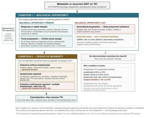
The two conditions are sequential decision domains rather than validated tests: the first concerns eligibility for benefit, the second feasibility of risk (Fig. 1). Both are required and, as Section 4.4 showed, they are not fully independent.
The two conditions governing operative selection in TKI-treated GIST. Abbreviations: TKI, tyrosine kinase inhibitor; ctDNA, circulating tumor DNA; SDH, succinate dehydrogenase; R0, microscopically complete resection. The first condition (biological opportunity) distinguishes states in which surgery could plausibly restore or prolong systemic control - response or stable disease, and focal progression with limited clonal escape - from generalized progression, where the opportunity is low. Liquid-biopsy evidence of multiple distinct secondary mutations appears as a marked investigational refinement and not as a decision criterion. The second condition (operative morbidity) grades the operation rather than the patient. The conditions are sequential decision domains, not validated binary tests, and they are not fully independent. The framework is hypothesis-generating and is not evidence of superiority over continued TKI therapy.
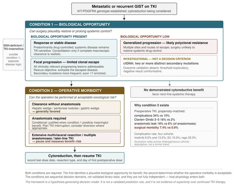
From kinase inhibition to earlier progression: the proposed causal chain. PDGFR inhibition leads to downregulation of Col1A1, Col3A1 and MMP2/9/12 at the anastomosis, thence to impaired anastomotic strength, leak, prolonged interruption of systemic therapy, and earlier progression. Solid boxes and arrows denote steps supported by the propensity-matched cohort, the histology, the animal model, or - for the terminal step - randomized evidence from BFR14, in which interruption of imatinib in controlled disease raised the risk of progression and of later resistance. The single dashed step, from complication to prolonged interruption, is near-mechanical but has never been quantified, since no cytoreduction series reports days to drug resumption. The residual uncertainty concerns duration rather than direction.
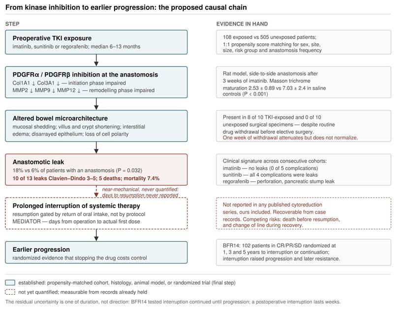
The escaped fraction as the boundary of surgical benefit. Left: focal progression, with a single escaped clone among lesions still controlled by drug; resection returns the patient to a drug-controllable state. Right: diffuse progression, with distinct secondary mutations at multiple sites; no set of resections restores control. Detection rates are lower bounds, since single-block sequencing cannot see heterogeneity it did not sample.
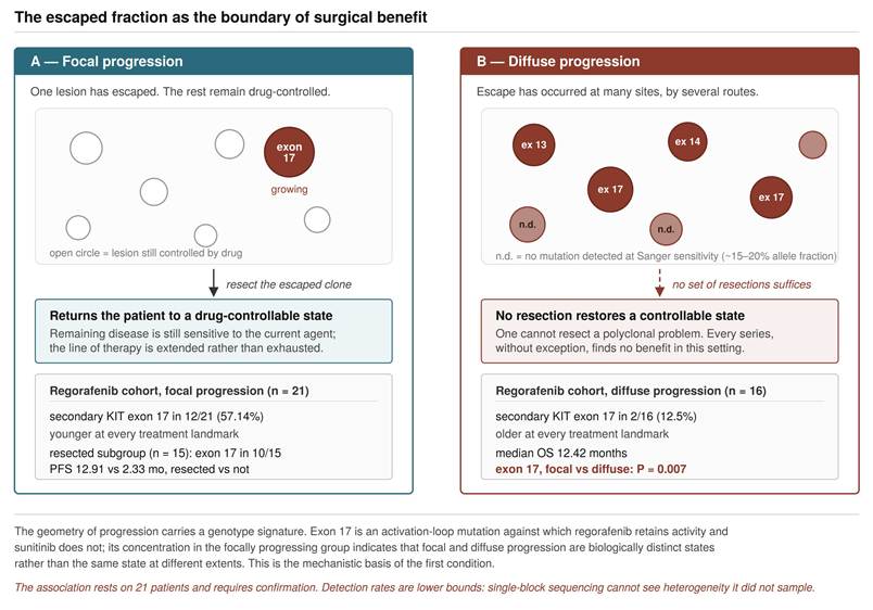
The first is passed by radiological response or stable disease, or by focal progression in which all clinically relevant progressing lesions can be addressed, and requires a known KIT/PDGFRA genotype; biological opportunity is low in generalized progression. The second is passed where clearance is achievable without anastomosis; conditionally where an anastomosis is required but justified by the first condition, with the interruption planned rather than defaulted; and should give pause where extensive multivisceral resection with multiple anastomoses is contemplated on a later-line agent.
The evidence supporting these settings is not equal, and the hierarchy should be stated rather than left implicit (Table 1). Focal progression in otherwise controlled disease is the best-supported indication, and it is also the setting in which the alternative is defined: not observation but the next line of therapy, whose median time to progression is known from registration trials [40, 41]. Consolidation of a response is plausible but unproven, and one finding constrains it. In BFR14, 74 patients reached complete response on imatinib alone with median overall survival of 154.1 months and 34 reached it with imatinib and resection of all lesions with 195.5 months, a difference that was not significant [24]. Conditional on reaching complete response the route did not detectably matter, and incomplete resection in that setting yielded survival close to that of not operating. In the consolidation setting, therefore, an operation should be undertaken only where conversion to complete response is realistic. That condition does not extend to rescue at focal progression, where the objective is extirpation of the escaped clone and gross clearance is achieved in only a small minority who nonetheless benefit [17].
Indications for cytoreduction in metastatic GIST, with comparator, appropriate endpoint and strength of supporting evidence.
| Setting | Objective of operation | Comparator | Appropriate endpoint | Evidence |
|---|---|---|---|---|
| Focal progression on any line | Extirpate the escaped fraction | Next-line TKI (registration-trial TTP) | PFS from operation vs next-line TTP | Best supported; level A + B |
| Response or stable disease | Convert to complete response | Continued TKI (counterfactual, unobservable) | Time to next-line TKI | Plausible, unproven; conditional on achievable R0 |
| Resection to forestall resistance | Reduce population at risk | Continued TKI | Time to secondary mutation | Hypothesis; premise supported, intervention untested |
| Generalized progression | None demonstrated | Next-line TKI | n/a | No demonstrated benefit; substantial morbidity |
| SDH-deficient / wild-type | Control symptomatic burden | Observation; TKI largely ineffective | Time to symptomatic burden | Separate disease-specific logic |
Perioperative morbidity after cytoreduction on TKI therapy, by line of therapy and by series.
| Series | Setting | n (patients unless stated) | Complications | Character of complications |
|---|---|---|---|---|
| Chang et al., 2015 | Imatinib, cytoreduction | 76 | 9.2% | Early and late groups comparable; two anastomotic leaks, both in the late group |
| Yeh et al., 2010 | Imatinib, PR/SD/LP | 38 ops | 13.2% | No anastomotic leak; wound, pulmonary and bleeding events |
| Yeh et al., 2017 | Sunitinib, focal progression | 26 | 15.3% | All four complications were anastomotic leaks; drug withdrawn 1 day |
| Yeh et al., 2021 | Regorafenib, focal progression | 15 | 33.3% | Perforation requiring reoperation; pancreatic stump leak; fatal recurrent GI bleeding |
| Raut et al., 2010 | Sunitinib, single center | 50 | 54% | 45 bowel resections yet no anastomotic leak tabulated; reoperation 16%; drug withdrawn 2-14 days |
| Fairweather et al., 2018 | Any TKI, two centers | 400 ops | 28% | Grade 3-5 after 18%; mortality 1.8%; 2 of 131 complications were leaks; drug withdrawn 1-14 days |
| Lee et al., 2019 | Any preoperative TKI (matched) | 108 | 34% | Grade 3-5 in 16%; anastomotic leak 18% of 71 anastomoses; surgical mortality 7.4% |
We state what this claims and what it does not. It proposes a framework for identifying patients in whom the expected benefit of an operation may plausibly exceed its drug-imposed morbidity, on the basis of consistent observational evidence, a molecular correlate of the selection variable, and a mechanistically characterized harm. It provides no numerical per-patient estimate of that difference, and the weighing it calls for is qualitative. It does not claim that cytoreduction improves survival relative to continued TKI therapy in a randomized comparison, because no such comparison exists and, for the reasons in Section 2, none is likely to be completed. Several components — the molecular correlate of response category, the exon 17 association, the impaired-healing mechanism, the graded complication profile — derive from consecutive cohorts at a single center, so external reproducibility is incomplete and the framework should be regarded as hypothesis-generating.
6. The comparator when no further systemic option remains
One assumption underlies the benchmark logic in the first condition and should be made explicit. Comparing an operation against the registration-trial time to progression of the next agent presumes that a next agent exists for that patient. Major guidelines direct patients who have exhausted approved therapy towards a clinical trial [7-9], which presumes an accessible trial. Where neither is available, the comparator is best supportive care rather than another drug, and the same observational estimate of surgical benefit is being weighed against a different alternative.
This is not a geographic argument and should not be read as one. Every patient with metastatic GIST who survives long enough reaches the end of the approved sequence, and one who has progressed through four lines and is eligible for no open protocol stands in the same position as a patient for whom the fourth line is not reimbursed. Variation in reimbursement timing and trial availability, which exists within Europe and North America as well as between continents, determines when that point is reached, not whether it is. Our third-line series was conducted when neither a fourth-line agent nor a trial of one was available locally [29], reported as a characteristic of the cohort rather than a justification of the practice.
The methodological consequence is familiar but has not been examined in this setting. The evidence on cytoreduction was generated where the next line is immediately available and evaluates surgery against the counterfactual of switching therapy. Where that counterfactual is absent the published estimate is a lower bound, and what is at issue is the transportability of the evidence, not its internal validity or the standard of proof required of it. Surgery here is best understood as a bridging strategy, holding disease until a further option arrives, in a landscape where options have continued to appear [42-44].
Two limits are stated as conditions rather than caveats. The absence of alternatives increases the expected value of an operation only among patients in whom the first condition is met; it does not create benefit where the biology provides none, nor license operating outside the framework. The patient who has exhausted every line is, by that fact, more likely to have a dominant resistant fraction — the state in which no series finds advantage and in which our data record 7.4% operative mortality. The temptation to operate because nothing else remains is therefore strongest where the first condition is most likely to fail, and constrained access argues for applying the second more strictly rather than less. Nothing here relaxes either condition.
7. What should be tested next
The field has repeatedly attempted to randomize the one question for which individual equipoise does not exist. Four questions retain it, given here in order of deliverability (Table 3).
Questions that retain equipoise, and analyses requiring no new patients.
| Question | Design | Why it is answerable |
|---|---|---|
| Perioperative TKI interruption duration | Randomized, 3 vs 7 vs 14 days, stratified by agent; endpoints anastomotic integrity, Clavien-Dindo >=III, days to resumption | Withholds no effective therapy; currently set by habit; deliverable in one high-volume center |
| Surgery vs next-line TKI at focal progression | Randomized; primary outcome time to subsequent progression | Genuine uncertainty; defined comparator; short accrual |
| Timing of resection relative to response plateau | Randomized in the neoadjuvant setting; endpoints organ preservation and R0 | Currently unspecified in every guideline |
| Cytoreduction vs continued current agent beyond progression | Randomized, where no further line is available | Control arm withholds nothing obtainable; cannot be run where the alternative exists |
| Complication to drug interruption to progression | Causal mediation analysis of existing records | No new patients; identifies the duration threshold BFR14 was not designed to find |
| Clonal architecture of focal progression | Multi-region sequencing of lesions resected at one operation | Banked tissue; tissue beats plasma because shedding is low |
| Burden-by-surgery interaction | Interaction term at a landmark in any cohort recording burden and operative status | Informative whichever way it resolves |
Perioperative TKI interruption duration is the most deliverable trial in this field and the practical endpoint of this review: randomization of drug-free interval, for example 3 versus 7 versus 14 days stratified by agent according to its elimination requirement, with anastomotic integrity, Clavien-Dindo [45] grade III or higher complications and days to resumption as endpoints. The question is currently answered by habit in every center, withholds no effective therapy, and is feasible in a single high-volume center or a small consortium.
Surgery versus immediate next-line therapy at focal progression retains genuine uncertainty, a defined comparator, and an outcome short enough to accrue. Timing of resection relative to the response plateau in the neoadjuvant setting is a third. A fourth applies wherever no further line is available: cytoreduction versus continuation of the current agent beyond progression, which withholds nothing the patient could otherwise receive and can only be conducted where no alternative agent exists.
Where randomization is not possible, the observational question should at least be posed as a trial that could in principle be run, and emulated against it. For cytoreduction in responsive metastatic disease that specification is: eligibility, metastatic KIT-mutant GIST with response or stable disease at six months of first-line imatinib and resectable residual burden; strategies, continued imatinib versus cytoreduction with protocol-defined perioperative interruption followed by resumption; time zero, the six-month landmark applied identically to both strategies rather than the date of operation; primary outcome, time to next-line therapy; safety co-primary, Clavien-Dindo grade III or higher and days to resumption. The landmark is illustrative rather than validated, chosen to fall beyond the usual plateau of imatinib response. Its importance is that almost every published series measures survival from the date of operation and thereby imports the entire interval from drug initiation to theatre as immortal time. Weighting on response depth, mutational subtype, site count and summed diameter addresses the measured confounders; an E-value should accompany the estimate.
Three analyses require no new patients. Mediation analysis of complication, days to resumption and progression-free survival from existing records would provide the first quantitative link between impaired healing and an oncological endpoint. Multi-region sequencing of lesions resected at one operation would convert a per-lesion detection rate into a per-patient clonal architecture; tissue rather than plasma is the substrate precisely because shedding is low. And the burden-by-surgery interaction predicted in Section 3.3 is testable in any cohort recording both.
8. What this review does and does not establish
Reviews of surgery in metastatic GIST have generally taken one of two forms: a chronological account of how practice changed after imatinib, or a summary of published series concluding that selected patients do well and that randomized data are awaited. Both are accurate and neither helps at the bedside, because both leave the operative decision where they found it. This review differs in taking the decision itself as the subject, and in accepting an obligation that the descriptive form avoids — to state, for each claim, what kind of evidence supports it.
Three positions distinguish it. The first is that response to TKI therapy is simultaneously a confounder and an effect modifier, and that the standard criticism of this literature errs not in identifying selection but in assuming the two roles are alternatives. The second is that the variable governing surgical benefit is the escaped fraction of tumor burden rather than the number of resistant clones, a reformulation adopted because the molecular heterogeneity data make any clone-counting framework untenable; we regard the earlier framing, including our own, as mistaken. The third is that the operative morbidity imposed by prior systemic therapy has an oncological price paid in interrupted drug exposure, and that this half of the decision has been described but never priced.
The review also disagrees with itself where the evidence does. We retract an interpretation we published in 2010; we identify a contradiction between our own timing data and the completeness requirement supported by BFR14, and resolve it by making that requirement intent-dependent; and having proposed that the preoperative drug-free interval explains the difference in reported anastomotic leak rates between centers, we withdraw that inference on finding that the published figures are not comparable quantities. Where a claim rests on 21 or 35 patients, we say so.
What the review does not do should be equally clear. It does not establish that cytoreduction improves survival over continued TKI therapy; no included study can support that, and the two trials designed to test it closed on accrual. It offers no numerical per-patient estimate of expected benefit against expected morbidity, and the weighing it proposes is qualitative. Several of its components derive from consecutive cohorts at a single center, so external reproducibility is incomplete and the framework should be treated as hypothesis-generating.
Its intended contribution is therefore modest and, we think, useful: a vocabulary for a decision currently made on undocumented individual thresholds, an explicit hierarchy of indications, a reporting standard for anastomotic complications that the field presently lacks, and four falsifiable propositions. If it prompts only the randomized comparison of perioperative interruption intervals — the one question here that is deliverable, withholds no effective therapy, and is currently answered by habit in every center performing this surgery — it will have served its purpose.
9. Conclusion
Surgery for GIST in the era of kinase inhibition is not a modality but a decision governed by two conditions. The first is biological: response identifies a clonal state that an operation can consolidate, focal progression identifies an escaped clone that an operation can remove, and generalized progression identifies a polyclonal process that no operation can address. The second is a morbidity condition the field has left largely unquantified: prolonged kinase inhibition alters the tissues on which the operation depends, at a price in complications, mortality and — the currency that matters oncologically — interrupted systemic therapy.
Selection remains a major limitation of this literature and no framework dissolves it. But selection does not by itself establish that the selected characteristic is clinically irrelevant: in GIST, response may simultaneously be a source of confounding and a biologically meaningful effect modifier, and a decision rule must accommodate both roles. What the literature most lacks is not randomization, which the structure of the disease will continue to withhold for the central question, but an accounting of both sides of the ledger and a willingness to randomize the questions that remain answerable. Of those, the perioperative interruption interval is the one that could be settled soonest.
Abbreviations
GIST: gastrointestinal stromal tumor; TKI: tyrosine kinase inhibitor; PFS: progression-free survival; OS: overall survival; RECIST: Response Evaluation Criteria in Solid Tumors; ctDNA: circulating tumor DNA; SDH: succinate dehydrogenase; CI: confidence interval; HR: hazard ratio.
AI usage statement
During the preparation of this manuscript, the authors used Claude (Anthropic) solely to revise wording and improve the clarity and readability of the English text. The AI tool was not used to design the manuscript structure, generate scientific content, select or interpret evidence, analyze data, or create figures. All AI-assisted wording changes were reviewed and edited by the authors, who take full responsibility for the content of the publication. AI tools are not listed as authors.
Competing Interests
The authors have declared that no competing interest exists.
References
1. Hirota S, Isozaki K, Moriyama Y. et al. Gain-of-function mutations of c-kit in human gastrointestinal stromal tumors. Science. 1998;279:577-580
2. DeMatteo RP, Lewis JJ, Leung D. et al. Two hundred gastrointestinal stromal tumors: recurrence patterns and prognostic factors for survival. Ann Surg. 2000;231:51-58
3. Joensuu H, Roberts PJ, Sarlomo-Rikala M. et al. Effect of the tyrosine kinase inhibitor STI571 in a patient with a metastatic gastrointestinal stromal tumor. N Engl J Med. 2001;344:1052-1056
4. Demetri GD, von Mehren M, Blanke CD. et al. Efficacy and safety of imatinib mesylate in advanced gastrointestinal stromal tumors. N Engl J Med. 2002;347:472-480
5. Fairweather M, Balachandran VP, Li GZ. et al. Cytoreductive surgery for metastatic gastrointestinal stromal tumors treated with tyrosine kinase inhibitors: a 2-institutional analysis. Ann Surg. 2018;268:296-302
6. Cavnar MJ, Seier K, Curtin C. et al. Outcome of 1000 patients with gastrointestinal stromal tumor (GIST) treated by surgery in the pre- and post-imatinib eras. Ann Surg. 2021;273:128-138
7. Koo DH, Ryu MH, Kim KM. et al. Asian consensus guidelines for the diagnosis and management of gastrointestinal stromal tumor. Cancer Res Treat. 2016;48:1155-1166
8. Casali PG, Blay JY, Abecassis N. et al. Gastrointestinal stromal tumours: ESMO-EURACAN-GENTURIS Clinical Practice Guidelines for diagnosis, treatment and follow-up. Ann Oncol. 2022;33:20-33
9. Hirano H, Naito Y, Takahashi T. et al. Updated Asian consensus guidelines for the diagnosis and management of gastrointestinal stromal tumor (2025). Gastric Cancer. 2026;29:887-903
10. Hirota S, Tateishi U, Nakamoto Y. et al. English version of Japanese Clinical Practice Guidelines 2022 for gastrointestinal stromal tumor (GIST) issued by the Japan Society of Clinical Oncology. Int J Clin Oncol. 2024;29:647-680
11. Du CY, Zhou Y, Song C. et al. Is there a role of surgery in patients with recurrent or metastatic gastrointestinal stromal tumours responding to imatinib: a prospective randomised trial in China. Eur J Cancer. 2014;50:1772-1778
12. Zaydfudim V, Okuno SH, Que FG. et al. Role of operative therapy in treatment of metastatic gastrointestinal stromal tumors. J Surg Res. 2012;177:248-254
13. An HJ, Ryu MH, Ryoo BY. et al. The effects of surgical cytoreduction prior to imatinib therapy on the prognosis of patients with advanced GIST. Ann Surg Oncol. 2013;20:4212-4218
14. Raut CP, Posner M, Desai J. et al. Surgical management of advanced gastrointestinal stromal tumors after treatment with targeted systemic therapy using kinase inhibitors. J Clin Oncol. 2006;24:2325-2331
15. DeMatteo RP, Maki RG, Singer S. et al. Results of tyrosine kinase inhibitor therapy followed by surgical resection for metastatic gastrointestinal stromal tumor. Ann Surg. 2007;245:347-352
16. Gronchi A, Fiore M, Miselli F. et al. Surgery of residual disease following molecular-targeted therapy with imatinib mesylate in advanced/metastatic GIST. Ann Surg. 2007;245:341-346
17. Yeh CN, Chen TW, Tseng JH. et al. Surgical management in metastatic gastrointestinal stromal tumor (GIST) patients after imatinib mesylate treatment. J Surg Oncol. 2010;102:599-603
18. Blanke CD, Rankin C, Demetri GD. et al. Phase III randomized, intergroup trial assessing imatinib mesylate at two dose levels in patients with unresectable or metastatic gastrointestinal stromal tumors expressing the KIT receptor tyrosine kinase: S0033. J Clin Oncol. 2008;26:626-632
19. Yeh CN, Chen YY, Tseng JH. et al. Imatinib mesylate for patients with recurrent or metastatic gastrointestinal stromal tumors expressing KIT: a decade experience from Taiwan. Transl Oncol. 2011;4:328-335
20. Antonescu CR, Besmer P, Guo T. et al. Acquired resistance to imatinib in gastrointestinal stromal tumor occurs through secondary gene mutation. Clin Cancer Res. 2005;11:4182-4190
21. Eisenhauer EA, Therasse P, Bogaerts J. et al. New response evaluation criteria in solid tumours: revised RECIST guideline (version 1.1). Eur J Cancer. 2009;45:228-247
22. Rutkowski P, Andrzejuk J, Bylina E. et al. What are the current outcomes of advanced gastrointestinal stromal tumors: who are the long-term survivors treated initially with imatinib? Med Oncol. 2013;30:765
23. Yeh CN, Wang SY, Tsai CY. et al. Surgical management of patients with progressing metastatic gastrointestinal stromal tumors receiving sunitinib treatment: a prospective cohort study. Int J Surg. 2017;39:30-36
24. Blay JY, Devin Q, Toulmonde M. et al. Twenty-year survival of advanced gastrointestinal stromal tumours treated with imatinib: exploratory long-term follow-up of the BFR14 trial. Ann Oncol. 2025;36:1035-1046
25. Chang SC, Liao CH, Wang SY. et al. Feasibility and timing of cytoreduction surgery in advanced (metastatic or recurrent) gastrointestinal stromal tumors during the era of imatinib. Medicine (Baltimore). 2015;94:e1014
26. Heinrich MC, Maki RG, Corless CL. et al. Primary and secondary kinase genotypes correlate with the biological and clinical activity of sunitinib in imatinib-resistant gastrointestinal stromal tumor. J Clin Oncol. 2008;26:5352-5359
27. Liegl B, Kepten I, Le C. et al. Heterogeneity of kinase inhibitor resistance mechanisms in GIST. J Pathol. 2008;216:64-74
28. Raut CP, Wang Q, Manola J. et al. Cytoreductive surgery in patients with metastatic gastrointestinal stromal tumor treated with sunitinib malate. Ann Surg Oncol. 2010;17:407-415
29. Yeh CN, Hu CH, Wang SY. et al. Cytoreductive surgery may be beneficial for highly selected patients with metastatic gastrointestinal stromal tumors receiving regorafenib facing local progression: a case controlled study. J Cancer. 2021;12:3335-3343
30. Rutkowski P, Bylina E, Klimczak A. et al. The outcome and predictive factors of sunitinib therapy in advanced gastrointestinal stromal tumors (GIST) after imatinib failure - one institution study. BMC Cancer. 2012;12:107
31. Heinrich MC, Jones RL, George S. et al. Ripretinib versus sunitinib in gastrointestinal stromal tumor: ctDNA biomarker analysis of the phase 3 INTRIGUE trial. Nat Med. 2024;30:498-506
32. Serrano C, Vivancos A, Lopez-Pousa A. et al. Clinical value of next generation sequencing of plasma cell-free DNA in gastrointestinal stromal tumors. BMC Cancer. 2020;20:99
33. Blay JY, Kang YK, Nishida T, von Mehren M. Gastrointestinal stromal tumours. Nat Rev Dis Primers. 2021;7:22
34. Blay JY, Devin Q, Duffaud F. et al. Discontinuation versus continuation of imatinib in patients with advanced gastrointestinal stromal tumours (BFR14): exploratory long-term follow-up of an open-label, multicentre, randomised, phase 3 trial. Lancet Oncol. 2024;25:1163-1175
35. Rutkowski P, Nowecki Z, Nyckowski P. et al. Surgical treatment of patients with initially inoperable and/or metastatic gastrointestinal stromal tumors (GIST) during therapy with imatinib mesylate. J Surg Oncol. 2006;93:304-311
36. Bauer S, Rutkowski P, Hohenberger P. et al. Long-term follow-up of patients with GIST undergoing metastasectomy in the era of imatinib - analysis of prognostic factors (EORTC-STBSG collaborative study). Eur J Surg Oncol. 2014;40:412-419
37. Lee JC, Chen CH, Chen TC, Yeh CN, Yeh TS. Preoperative tyrosine kinase inhibitors risks bowel anastomotic healing in patients with advanced primary and recurrent/metastatic gastrointestinal stromal tumors - a rose has its thorns. Eur J Surg Oncol. 2019;45:153-159
38. Rajkumar VS, Shiwen X, Bostrom M. et al. Platelet-derived growth factor-beta receptor activation is essential for fibroblast and pericyte recruitment during cutaneous wound healing. Am J Pathol. 2006;169:2254-2265
39. Shah DR, Dholakia S, Shah RR. Effect of tyrosine kinase inhibitors on wound healing and tissue repair: implications for surgery in cancer patients. Drug Saf. 2014;37:135-149
40. Demetri GD, van Oosterom AT, Garrett CR. et al. Efficacy and safety of sunitinib in patients with advanced gastrointestinal stromal tumour after failure of imatinib: a randomised controlled trial. Lancet. 2006;368:1329-1338
41. Demetri GD, Reichardt P, Kang YK. et al. Efficacy and safety of regorafenib for advanced gastrointestinal stromal tumours after failure of imatinib and sunitinib (GRID): an international, multicentre, randomised, placebo-controlled, phase 3 trial. Lancet. 2013;381:295-302
42. Blay JY, Serrano C, Heinrich MC. et al. Ripretinib in patients with advanced gastrointestinal stromal tumours (INVICTUS): a double-blind, randomised, placebo-controlled, phase 3 trial. Lancet Oncol. 2020;21:923-934
43. Heinrich MC, Jones RL, von Mehren M. et al. Avapritinib in advanced PDGFRA D842V-mutant gastrointestinal stromal tumour (NAVIGATOR): a multicentre, open-label, phase 1 trial. Lancet Oncol. 2020;21:935-946
44. Heinrich MC, Zhang X, Jones RL. et al. Clinical benefit of avapritinib in KIT-mutant gastrointestinal stromal tumors: a post hoc analysis of the phase I NAVIGATOR and phase I/II CS3007-001 studies. Clin Cancer Res. 2024;30:719-728
45. Dindo D, Demartines N, Clavien PA. Classification of surgical complications: a new proposal with evaluation in a cohort of 6336 patients and results of a survey. Ann Surg. 2004;240:205-213
Author contact
Corresponding author: Chun-Nan Yeh, MD, Professor of Surgery, Department of Surgery, Chang Gung Memorial Hospital, 5 Fu-Hsing Street, Kwei-Shan, Taoyuan, Taiwan. E-mail: yehchunnancom.