Impact Factor ISSN: 1837-9664




J Cancer 2026; 17(12):2082-2102. doi:10.7150/jca.141758 This issue Cite
Review
The Roles of Vitamins in Cancer Initiation, Progression, and Therapy: Clinical Evidence, Molecular Mechanisms, and Precision Translation
Fanglin Liu1,2#, Anqi He1#, Liang Zhou1, Yong Xia1  , Xuyi Wu1 , Fuyan Xu3
1. Rehabilitation Medicine Center and Institute of Rehabilitation Medicine, Rehabilitation Medicine Key Laboratory of Sichuan Province, West China Hospital, Sichuan University, Chengdu, 610041, China.
2. West China School of Pharmacy, Sichuan University, Chengdu 610041, China.
3. Joint Lab of Reproductive Medicine of SCU-CUHK, Lab of Reproductive genetics and Epigenetics, Department of Obstetrics/Gynecology, Key Laboratory of Birth Defects and Related Disease of Women and Children of MOE, West China Second University Hospital, Sichuan University, 610041, Chengdu, China.
# These authors contributed equally to this work.
Received 2026-7-30; Accepted 2026-10-28; Published 2026-11-13
Abstract
Vitamins are essential micronutrients required for normal physiology, yet their roles in cancer may extend far beyond basic nutritional support. Increasing evidence suggests that vitamins could influence cancer initiation, progression, metastasis, therapeutic response, and survivorship through highly context-dependent mechanisms. Epidemiological and clinical studies indicate that adequate vitamin status may be associated with reduced incidence of selected cancers and improved post-diagnosis outcomes, particularly for vitamin D in colorectal and breast cancers. However, these associations are heterogeneous, and high-dose or inappropriate supplementation may be ineffective or even unfavorable in specific cancer types, molecular contexts, or treatment settings. Mechanistically, in preclinical studies, vitamins could regulate tumor biology through nuclear receptor signaling, epigenetic and RNA modification, redox homeostasis, DNA damage responses, ferroptosis, metabolic reprogramming, inflammatory signaling, and tumor cell death. Recent studies further highlight emerging mechanisms, including vitamin availability and transporter dependency, vitamin-derived protein modifications, microbiota-mediated vitamin D metabolism, and competition for vitamins between tumor and immune cells. In the tumor microenvironment, vitamins could reshape gut barrier function, antigen presentation, myeloid cell states, CD8⁺ T-cell fitness, natural killer (NK)-cell activity, vascular normalization, and metastatic niches. Clinically, vitamins are being explored as adjuncts to chemotherapy, radiotherapy, immunotherapy, targeted therapy, and supportive care, with high-dose intravenous vitamin C, vitamin D, and selected B vitamins receiving particular attention. This review summarizes current clinical evidence, mechanistic insights, therapeutic implications, and ongoing controversies, and emphasizes the need for precision vitamin-based strategies guided by cancer type, baseline vitamin status, dose, route, timing, molecular subtype, and immune context.
Keywords: vitamins, carcinogenesis, cancer therapy, cancer immunity, metabolic reprogramming, epigenetic regulation, clinical translation
Introduction
Cancer initiation, progression, metastasis, and therapeutic response are shaped by genetic alterations, epigenetic dysregulation, metabolic reprogramming, chronic inflammation, immune escape, and remodeling of the tumor microenvironment. As oncology has shifted from a cancer cell-centered model toward an integrated “tumor-host-microenvironment” view, increasing attention has focused on how host nutritional status, diet, metabolic homeostasis, and the microbiome influence the cancer continuum. Vitamins, as essential micronutrients, have traditionally been regarded as nutrients required for development, tissue homeostasis, and basal metabolism. Accumulating evidence now shows that they also regulate cell proliferation, differentiation, death, redox balance, DNA damage repair, inflammation, and immunity, thereby influencing cancer risk, progression, and treatment sensitivity [1-3].
Unlike general nutritional support, the roles of vitamins in cancer are complex and context-dependent. Vitamin deficiency or metabolic dysregulation may impair tissue homeostasis, weaken immune surveillance, promote chronic inflammation, and increase genomic instability, creating conditions favorable for tumor initiation and progression. However, more is not necessarily better. In specific cancer types, molecular backgrounds, disease stages, dose ranges, or treatment settings, excessive exposure or inappropriate supplementation may provide little benefit, support tumor metabolic demands, alter therapeutic response, or interfere with antitumor immunity [4-8]. Recent studies further suggest that vitamins can serve as contested metabolic resources between tumor and immune cells. A preclinical study found that pancreatic cancer cells can consume vitamin B6 to support one-carbon metabolism and suppress NK-cell function [9], whereas vitamin B5 supports MYC-driven coenzyme A metabolism and metabolic adaptation in breast cancer [10]. Thus, vitamins should not be viewed simply as nutritional supplements, but as bioactive regulators of tumor metabolic ecology.
Current research on vitamins and cancer can be divided into three areas. First, epidemiological and clinical studies examine associations between vitamin intake, circulating levels, supplement use, cancer incidence, and post-diagnosis survival. For example, low plasma vitamin D is associated with poorer survival after colorectal cancer surgery [11], while sufficient vitamin D levels are linked to improved survival and cardiometabolic outcomes in breast cancer survivors [12]. Second, clinical and preclinical studies are testing vitamins as adjunctive interventions in chemotherapy, radiotherapy, immunotherapy, targeted therapy, and symptom management. Third, mechanistic studies indicate that vitamins influence both tumor-intrinsic processes and the tumor microenvironment (TME). In addition to metabolic and epigenetic regulation [13, 14], primary studies implicate vitamin D-VDR signaling in intestinal barrier homeostasis and microbiota-dependent cancer immunity [15, 16], while vitamins C and E can modulate dendritic-cell function and subsequent T-cell responses [17-19].
Despite these advances, substantial heterogeneity remains. Different vitamins act through distinct mechanisms, and the same vitamin may have opposite effects across cancer types, doses, and treatment stages. Observational studies, randomized trials, and preclinical models are not always consistent, and physiological supplementation should be distinguished from pharmacological intervention. Therefore, this review summarizes current evidence on vitamins in cancer incidence, survival, treatment response, symptom control, tumor-intrinsic mechanisms, immune regulation, and ongoing clinical trials, aiming to support precise, stratified, and mechanism-guided use of vitamins in cancer prevention and therapy.
Clinical impact of vitamins on cancer incidence and post-diagnosis survival
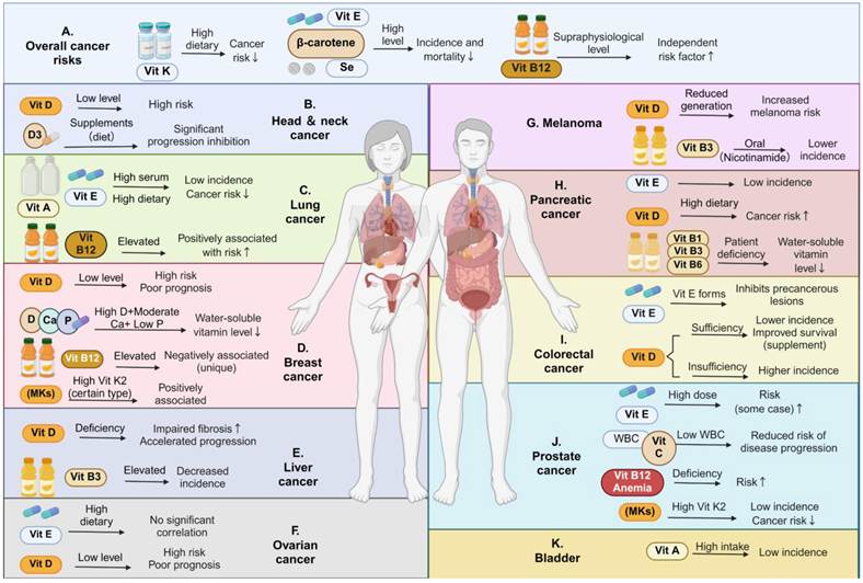
Dietary components and lifestyle factors (such as exercise) are important factors influencing cancer incidence [20]. Diet is an important source of vitamins in the body. The relationship between vitamins, cancer incidence, and post-diagnosis survival is one of the most clinically studied but heterogeneous areas in vitamin oncology. Current evidence mainly examines dietary intake, supplement use, and circulating levels. Overall, adequate vitamin status may be associated with lower risk of selected cancers and better survival, but these effects are not universal and do not imply that high-dose supplementation is necessarily beneficial. Cancer type, exposure source, dose, baseline nutritional status, and disease stage all shape the clinical relevance of vitamins. Figure 1 summarizes the major clinical associations between vitamins and cancer incidence, while Table 1 provides representative evidence.
The multifaceted roles of vitamins and minerals in cancer incidence, progression, and prognosis. (A) Systemic effects on overall cancer risk: Higher dietary intake of vitamin K, vitamin E, β-carotene, and selenium is generally associated with reduced cancer incidence and mortality, while supraphysiological circulating levels of vitamin B₁₂ may function as an independent risk predictor. (B-K) Organ-specific impacts of micronutrient status: The central schematic demonstrates the influence of vitamins A, B-complex, C, D, E, K, and essential minerals (calcium, phosphorus) across 10 common cancer types. Abbreviations. Vit: vitamin; Vit A: vitamin A; Vit B1: thiamine; Vit B3: niacin; Vit B6: pyridoxine; Vit B12: cobalamin; Vit C: vitamin C; Vit D: vitamin D; D3: vitamin D3 (cholecalciferol); Vit E: vitamin E; Vit K: vitamin K; Vit K2: vitamin K2 (menaquinones); MKs: menaquinones; Se: selenium; Ca: calcium; P: phosphorus; WBC: white blood cells.
A summary of clinical evidence regarding the impact of vitamins on cancer incidence and post-diagnosis survival rates
| Vitamin | Major types of cancer | Exposure indicators | Key clinical findings | Types of evidence | References |
|---|---|---|---|---|---|
| Vitamin D | Oral cancer | Vitamin D deficiency, VDR expression/polymorphism, vitamin D-related interventions | Vitamin D deficiency is associated with an increased risk of oral cancer; VDR polymorphisms or abnormal expression may be associated with an increased risk and poorer survival, although some studies have not found a clear association. | Scoping review. Included in case-control studies, cohort studies, and clinical trials. | [21] |
| Vitamin D | Lung cancer | Pre-diagnostic serum 25(OH)D levels | No clear association was found between circulating 25(OH)D levels and the risk of lung cancer. | A nested case-control/consortium analysis of 20 prospective cohorts | [22] |
| Vitamin D | Colorectal cancer | Serum 25(OH)D levels | Higher serum 25(OH)D levels are associated with a reduced risk of early- and late-onset colorectal cancer, with the association being more pronounced in younger individuals. | Large-scale population cohort study | [23] |
| Vitamin D | All-cancer mortality | Vitamin D3 supplement, 2,000 IU/day | Vitamin D supplementation did not significantly reduce the overall incidence of invasive cancers; while there was a trend toward reduced cancer mortality, it did not reach statistical significance for the primary endpoint. | Randomized, placebo-controlled clinical trial | [24] |
| Vitamin D | All-cancer mortality | Vitamin D3 supplements, 1,600 IU/day or 3,200 IU/day | Among older adults with generally adequate baseline vitamin D levels, vitamin D supplementation did not reduce the incidence of invasive cancers | Randomized, placebo-controlled clinical trial | [25] |
| Vitamin D | Various cancers and cancer mortality | Serum 25(OH)D Levels and Vitamin D Supplementation | Observational studies generally support a negative association between serum 25(OH)D levels and the incidence, metastasis, and mortality of various cancers. However, some large-scale RCTs have failed to confirm a reduction in cancer incidence. | Systematic Review | [26] |
| Vitamin D | All cancers, prostate cancer, breast cancer, colorectal cancer, lung cancer, melanoma | Vitamin D3 supplement, 60,000 IU per month | No evidence was found that vitamin D supplementation reduces the overall incidence of cancer or the incidence of major individual cancer types. Its potential impact on cancer mortality may be more closely related to survival. | Randomized, placebo-controlled clinical trial | [27] |
| Vitamin D | Overall cancer incidence and cancer mortality | Vitamin D3 supplement, 2,000 IU/day | No significant reduction in the overall incidence of invasive cancers; an updated meta-analysis suggests a possible reduction in cancer mortality rather than cancer incidence | RCTs and Updated Meta-Analyses | [28] |
| Vitamin D | Squamous cell carcinoma of the head and neck | Serum 25(OH)D Levels and Psychological Distress | Vitamin D deficiency demonstrated a non-significant trend toward association with poorly differentiated tumors; after adjusting for confounding factors, it did not independently predict poor differentiation. | Cross-sectional clinical study | [29] |
| Vitamin D | Skin Melanoma | High-dose vitamin D supplementation, 100,000 IU per month | Vitamin D supplementation increases serum 25(OH)D levels but does not improve recurrence-free survival, melanoma-related mortality, or overall survival | Randomized, double-blind, placebo-controlled trial | [30] |
| Vitamin D | Gastrointestinal Cancers | Vitamin D3 supplementation, 2000 IU/day; p53 immune response status | In the subgroup of gastrointestinal cancers with positive p53 immune responses, vitamin D supplementation reduced the risk of recurrence or death; no benefit was observed in the subgroup without positive p53 immune responses. | Post-hoc Subgroup Analysis of the AMATERASU Trial | [31] |
| Vitamin D | Colorectal cancer | Postoperative plasma 25(OH)D levels | Higher postoperative 25(OH)D levels are associated with lower rates of colorectal cancer-specific mortality and all-cause mortality, particularly among stage II patients. | Prospective cohort study | [32] |
| Vitamin D | Metastatic colorectal cancer | High-dose versus standard-dose vitamin D3 supplementation | The improvement in median PFS in the high-dose group did not reach conventional statistical significance, but a supportive Cox model suggests a reduced risk of PFS or death; OS was not significantly improved. | Randomized Phase II clinical trial | [33] |
| Vitamin D | Colorectal cancer | Vitamin D supplementation | Vitamin D supplementation is associated with a reduced risk of adverse outcomes in colorectal cancer and may improve progression-free survival or cancer-specific survival | Systematic Reviews and Meta-Analyses of RCTs | [34] |
| Vitamin A | Bladder cancer | Serum vitamin A levels, retinoic acid signaling, and gut microbiota-related markers | Patients with bladder cancer may exhibit reduced serum vitamin A levels and an imbalance in the gut microbiota; the vitamin A-gut microbiota-retinoic acid (RA) signaling pathway may be associated with bladder cancer prevention | Review | [35] |
| Vitamin A | Prostate cancer | Serum retinol, lutein, α-carotene, β-carotene, etc. | Various forms of vitamin A and carotenoids are associated with an increased risk of prostate cancer, and this association is influenced by smoking and alcohol consumption. | Hospital Case-Control Study | [36] |
| Vitamin E | Prostate cancer | Serum α-tocopherol, δ/γ/α- tocotrienol, etc. | Various forms of vitamin E are associated with an increased risk of prostate cancer, and this association is influenced by smoking and alcohol consumption | Hospital Case-Control Study | [36] |
| Vitamin E | Various types of cancer | Tocotrienol-type vitamin E analogues | Tocotrienols have shown potential in preventing or inhibiting tumor growth in various types of cancer, but evidence from clinical incidence remains limited. | Review covering in vitro, animal, and clinical studies | [37] |
| Vitamin E / Vitamin C / Selenium | Gastric cancer | Combined supplementation with vitamin C, vitamin E, and selenium | Long-term supplementation with a combination of vitamins is associated with a reduced incidence of gastric cancer and a lower risk of death from gastric cancer | Long-term follow-up of randomized, placebo-controlled intervention trials | [38] |
| Vitamin E | Prostate cancer | Dietary vitamin E intake, vitamin E supplement intake | Overall, dietary or supplemental vitamin E intake was not significantly associated with prostate cancer risk; a protective trend may exist in certain subgroups. | Meta-analysis | [39] |
| Vitamin E / Selenium | High-grade prostate cancer | Vitamin E/selenium supplementation and mutations in related metabolic/transport genes | Variants in genes related to vitamin E or selenium metabolism and transport may influence the risk of overall and high-grade prostate cancer and alter an individual's response to supplements | Genetic analysis of the SELECT case cohort | [40] |
| Vitamin E/β-carotene | Prostate cancer | Alpha-tocopherol and beta-carotene supplements | Among male smokers, α-tocopherol supplementation reduces the incidence and mortality of prostate cancer; β-carotene has not been shown to provide protection, and there is a trend toward increased incidence and mortality, though this is not definitive. | Randomized controlled trial | [41] |
| Vitamin K | Various types of cancer | Vitamin K supplementation or exposure to vitamin K | Vitamin K may exert anticancer effects through anti-inflammatory, antioxidant, anti-aging, and anti-metastatic mechanisms; however, clinical evidence regarding incidence and survival remains insufficient. | Review | [42] |
| Vitamin K | Lung cancer | Dietary vitamin K intake | Vitamin K intake is inversely associated with the risk of lung cancer, and this association is more pronounced among men and former smokers. | Large-scale prospective cohort study | [43] |
| Vitamin K | Cancer Cachexia and Death | Dietary vitamin K intake | Vitamin K intake is inversely associated with the incidence of cancer cachexia; high intake is associated with a reduced risk of all-cause mortality and cardiovascular mortality among survivors of cancer cachexia. | NHANES cohort analysis | [44] |
| Vitamin B12 | Hospitalized patients with various types of cancer | Plasma vitamin B12 levels | Abnormal vitamin B12 levels are common in cancer patients; elevated B12 levels are associated with poorer clinical status, advanced disease, and liver failure, while B12 deficiency is more common in patients with early-stage cancer and the elderly. | Retrospective clinical study | [45] |
| Vitamin B1/B2/B6 | Gastric cancer | Dietary intake of vitamins B1, B2, and B6 | Higher dietary intakes of vitamins B1 and B6 are associated with a reduced risk of gastric cancer; vitamin B2 intake is not significantly associated with gastric cancer risk. | Hospital case-control study | [46] |
| Vitamin B6/B12 | Pancreatic cancer | Vitamin B6/B12 intake, blood PLP/B12 levels | High vitamin B6 intake and high blood PLP levels are associated with a reduced risk of pancreatic cancer; vitamin B12 intake or blood levels are not significantly associated with pancreatic cancer risk | Meta-analysis | [47] |
| Vitamin B2 | Colorectal cancer | Dietary/total vitamin B2 intake, blood vitamin B2 levels | Vitamin B2 intake is associated with a reduced risk of colorectal cancer; higher blood levels of vitamin B2 may also be associated with a reduced risk | Dose-response Meta-analysis | [48] |
| Vitamin C | Cancers of the digestive system | Dietary vitamin C intake, plasma vitamin C levels | Dietary vitamin C intake is associated with a reduced overall risk of digestive system cancers, particularly cancers of the mouth, pharynx, and esophagus, as well as gastric and colon cancers; plasma vitamin C levels are significantly associated only with a reduced risk of gastric cancer | Systematic reviews and meta-analyses of prospective studies | [49] |
| Vitamin C | Endometrial cancer | Genetically predicted circulating vitamin C levels | Genetically predicted vitamin C levels are associated with an increased risk of endometrial cancer, suggesting that this effect may be cancer-specific | Mendelian randomization study | [50] |
Abbreviations: PFS: progression-free survival; OS: overall survival; 25(OH)D: 25-hydroxyvitamin D.
Vitamin D
Vitamin D is the most extensively studied vitamin in cancer, and serum 25(OH)D is the main marker of vitamin D status. Low 25(OH)D or vitamin D deficiency has been associated with increased risk of colorectal, breast, ovarian, head and neck, and prostate cancers [21, 23, 26]. Evidence is strongest for colorectal cancer. In observational studies, higher serum 25(OH)D is associated with lower risk of both early- and late-onset colorectal cancer (CRC) [23], and higher total vitamin D intake is linked to reduced risk of early-onset CRC and its precursors [51]. After diagnosis, low plasma 25(OH)D is associated with poorer survival after CRC surgery, independent of systemic inflammation [11]. In breast cancer survivors, sufficient vitamin D levels are also associated with better overall survival, disease-free survival, and second primary cancer-free survival [12]. However, vitamin D is not a broad-spectrum cancer-preventive factor. Large randomized trials in general populations have not shown a significant reduction in overall invasive cancer incidence [24, 25, 27], and prospective lung cancer analyses found no clear association between 25(OH)D and lung cancer risk [22]. Thus, vitamin D may be most relevant in deficient individuals, selected cancer types, or specific molecular contexts.
B Vitamins
B vitamins show more bidirectional associations because they are deeply involved in one-carbon metabolism, nucleotide synthesis, methyl donor supply, and energy metabolism. Higher vitamin B1 and B6 intake has been associated with lower gastric cancer risk [46], higher vitamin B6 intake or blood pyridoxal 5′-phosphate (PLP) levels with lower pancreatic cancer risk [47], and higher vitamin B2 intake or blood levels with lower CRC risk [48]. In contrast, evidence for vitamin B12 is more complex. Elevated serum B12 has been associated with higher risk of lung, liver, pancreatic, and some hematological cancers [45, 52]. However, this does not necessarily indicate direct carcinogenicity. High B12 may also reflect liver dysfunction, inflammation, tumor burden, or altered binding proteins. Therefore, interpretation of B vitamins requires distinguishing dietary intake, supplement use, abnormal circulating levels, and functional deficiency.
Vitamin C
Clinical evidence on vitamin C and cancer incidence mainly concerns digestive system cancers. A recent systematic review and meta-analysis of 32 prospective studies showed that higher dietary vitamin C intake was associated with lower overall digestive cancer risk, especially oral/pharyngeal/esophageal, gastric, and CRC; however, plasma vitamin C was significantly associated only with lower gastric cancer risk [49]. Combined supplementation with vitamin C, vitamin E, and selenium also reduced gastric cancer incidence and mortality during long-term follow-up in a high-risk population [38]. Nevertheless, vitamin C is not uniformly protective. A Mendelian randomization study suggested that genetically predicted circulating vitamin C may be associated with higher endometrial cancer risk [50]. Evidence supporting vitamin C as an independent prognostic marker remains limited, and its clinical meaning may largely reflect oxidative stress, inflammatory burden, and nutritional reserve.
Vitamins A, E, and K
Clinical evidence for vitamins A, E, and K is more scattered and highly cancer- and population-specific. Vitamin A and its related retinol, carotenoids and retinoic acid participate in epithelial differentiation and mucosal homeostasis and may be relevant to bladder cancer prevention [35]. However, higher serum retinol, α-carotene, and β-carotene may be associated with increased prostate cancer risk [36]. For vitamin E, higher dietary intake has been linked to reduced risk of some cancers [53], but its protective effect in prostate cancer is inconsistent and may depend on vitamin E isoform, supplement dose, smoking status, and genetic background [36, 39-41]. Evidence for vitamin K is limited. Dietary vitamin K intake is associated with lower lung cancer risk [43], but its relationships with cancer cachexia, survival, and some breast cancer subtypes may be nonlinear [44, 54].
Overall, the associations between vitamins, cancer incidence, and post-diagnosis survival are not uniform. The same vitamin may be protective, neutral, or adverse across different cancers. Circulating vitamin levels may participate in disease biology, but may also mark tumor burden, inflammation, organ dysfunction, or nutritional status. Future studies should stratify by cancer type, disease stage, baseline vitamin status, genetic background, and treatment context to identify patients most likely to benefit from vitamin optimization.
Effects of vitamins on cancer treatment efficacy and cancer-related symptoms
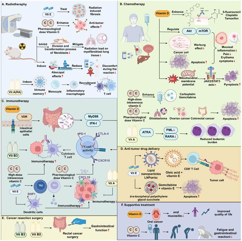
Beyond cancer incidence and prognosis, vitamins may alter tumor sensitivity to existing therapies and affect toxicity and symptoms. These effects differ from routine nutritional supplementation and depend on dose, route, combination strategy, and patient selection. Figure 2 summarizes the potential roles of vitamins in chemotherapy, radiotherapy, immunotherapy, targeted therapy, and supportive care.
Synergistic roles of vitamins in multimodal cancer therapy and supportive care. (A) Radiotherapy: Vitamin E reduces radiation-induced fibrosis. High-dose vitamin C enhances antitumor effects and inhibits lung tissue remodeling. Vitamin A induces immune memory and promotes immune cell infiltration, strengthening abscopal-like effects. (B) Chemotherapy: Vitamin D synergizes with chemotherapeutics via Akt/mTOR and JAK2/STAT3 signaling to inhibit the Warburg effect and induce apoptosis. High-dose intravenous Vitamin C boosts the efficacy of multiple chemotherapies in solid tumors. Vitamin A treats acute promyelocytic leukemia by promoting PML/RARA degradation. (C) Immunotherapy: Vitamin D regulates the intestinal immune axis. Vitamins B3, C and E enhance T-cell and DC function, while high-dose vitamin C promotes T-cell infiltration to improve PD-1/CTLA-4 blockade efficacy. (D) Drug delivery: Vitamin E derivatives in LNPs improve chemotherapeutic delivery and CD8⁺ T-cell infiltration. (E, F) Surgery and supportive care: Perioperative B vitamins accelerate postoperative recovery. Vitamins D and C alleviate mucositis and fatigue, improving quality of life. Abbreviations. Vit: vitamin; Vit A: vitamin A; Vit B3: vitamin B3; Vit B5: vitamin B5; Vit E: vitamin E; RA: retinoic acid; PML: promyelocytic leukemia protein; ATRA: All-trans retinoic acid; RARA: retinoic acid receptor alpha; PD-1: programmed cell death protein 1; CTLA-4: cytotoxic T-lymphocyte-associated protein 4; CXCR6: C-X-C motif chemokine receptor 6; CXCL16: C-X-C motif chemokine ligand 16; Th1: type 1 helper T cells; Th2: type 2 helper T cells; SHP1: Src homology 2 domain-containing protein tyrosine phosphatase 1; LNP: lipid nanoparticle; LNP(OP/EC): lipid nanoparticle formulation combining oleic acid-scaffold and vitamin E-scaffold ionizable lipids.
Chemotherapy
Vitamin D has received considerable attention. In metastatic CRC, high-dose vitamin D3 combined with mFOLFOX6 and bevacizumab showed a trend toward improved PFS compared with standard-dose vitamin D3, and a supportive Cox model suggested reduced risk of progression or death [33]. In breast cancer receiving neoadjuvant systemic therapy, oral vitamin D supplementation was associated with a higher pathological complete response rate [55]. Vitamin C is closer to a pharmacological adjuvant. Because oral dosing cannot achieve millimolar plasma concentrations, high-dose intravenous vitamin C has become the main clinical strategy [56-58]. In metastatic pancreatic ductal adenocarcinoma (PDAC), pharmacological ascorbate plus gemcitabine and nab-paclitaxel prolonged overall and progression-free survival without worsening quality of life or severe toxicity [59]. Preclinical studies show that 1,25-dihydroxyvitamin D3 enhances cisplatin sensitivity in bladder cancer cells by suppressing the Warburg effect through modulation of AKT/mTOR signaling [60]. Pharmacological ascorbate also enhances the antitumor effects of carboplatin and paclitaxel in ovarian cancer mouse models [61].
Radiotherapy
In radiotherapy, vitamins may provide both radiosensitization and normal tissue protection. Clinically, oral pentoxifylline and vitamin E may alleviate radiation-induced fibrosis, particularly in breast and head-and-neck cancer survivors [62]. Pharmacological vitamin C has also been explored for reducing radiation-related fatigue, gastrointestinal reactions, and tissue injury [63]. Preclinical studies show that high-dose vitamin C can enhance radiation-induced tumor suppression while inhibiting fibroblast-to-myofibroblast transition and reducing radiation-induced pulmonary fibrosis [64]. RA combined with local radiotherapy can also induce abscopal effects and immune memory, suggesting that vitamin A derivatives may help convert local irradiation into a systemic antitumor immune event [65]. Thus, vitamin use during radiotherapy should be evaluated for both tumor control and tissue protection, rather than judged solely by antioxidant activity.
Immunotherapy
The impact of vitamins on immunotherapy mainly reflects remodeling of the tumor immune microenvironment. Clinically, the prospective PROVIDENCE study showed that systematic vitamin D supplementation in patients with advanced cancer receiving immune checkpoint inhibitors was associated with longer overall survival, longer time to treatment failure, higher disease control rate, and lower risk of thyroid immune-related adverse events [66]. This finding may suggest a potential favorable effect of vitamin D on the efficacy of immune checkpoint inhibitors; however, given the observational design of PROVIDENCE, this association cannot be interpreted as causal, and confirmation in randomized controlled trials is warranted. Mechanistic studies further show that vitamin D can regulate microbiome-dependent antitumor immunity and may affect immunotherapy response [15]. Vitamin C promotes CD8⁺ T-cell infiltration, interferon (IFN)-related signaling, and stimulator of interferon Genes (STING) activation in specific preclinical models [67-70]. Magrì et al. further demonstrated that high-dose vitamin C enhances the antitumor activity of anti-PD-1 and anti-CTLA-4 therapy in mouse cancer models. The combination increased intratumoral T-cell infiltration and activation, and produced durable complete responses with antitumor immune memory in selected mismatch repair-deficient models [18]. Vitamin B3 relieves myeloid immunosuppression and enhances CD8⁺ T-cell effector function [71]. Vitamin E inhibits Src homology 2 domain-containing protein tyrosine phosphatase 1 (SHP1) in dendritic cells, thereby improving antigen presentation and T-cell priming [17]. These data suggest that vitamins may act as immune-ecological modulators rather than simple immune stimulants.
Targeted therapy
Resistance and dose-limiting toxicity remain major obstacles to targeted therapy. Vitamins may enhance sensitivity by reshaping metabolism, epigenetics, and the immune microenvironment. In liver cancer, vitamin B3 inhibits nuclear factor kappa-light-chain-enhancer of activated B cells (NF-κB) activation through G-protein-coupled receptor 109A (GPR109A) /β-arrestin signaling, relieves myeloid immunosuppression, and enhances lenvatinib activity [71]. In triple-negative breast cancer, vitamin C enhances the efficacy of BET and PI3K inhibitors in small-sample/preclinical studies, although these findings require validation in larger multicenter randomized trials [72, 73]. In chronic lymphocytic leukemia, ascorbate synergizes with ibrutinib, idelalisib, and venetoclax and partly overcomes protection mediated by bone marrow stroma, cytokines, and hypoxia [74]. Vitamin C has also been reported to restrict acquired resistance to EGFR-targeted therapy in colorectal cancer [75].
Other treatment strategies
Vitamin derivatives can also function as direct therapeutic strategies in selected cancers. All-trans retinoic acid (ATRA) degrades PML-RARA and relieves differentiation arrest, making it a standard component of therapy for acute promyelocytic leukemia [76]. In metastasis control, a stable vitamin C derivative suppresses renal cancer bone metastasis by reducing stem-like features, cell-cycle progression, osteoclast differentiation, and neutrophil recruitment; combination with a CXCR2 antagonist further inhibits bone metastasis [77]. These findings suggest roles for vitamin-based approaches in differentiation therapy, metastatic niche remodeling, and relapse prevention.
Cancer-related symptoms, treatment toxicity, and quality of life
Vitamins may also improve symptoms and treatment tolerance by correcting deficiencies, reducing inflammation, and supporting tissue repair. Vitamin D is closely linked to bone metabolism, muscle function, pain, and infection risk; in palliative care, supplementation may reduce pain and infection incidence [78]. In brain tumor resection patients with low vitamin D levels, a single preoperative high-dose vitamin D3 injection shortened ICU and hospital stay [79]. A small-sample preliminary study showed that vitamin may relieve fatigue, gastrointestinal discomfort, and inflammation-related symptoms, and high-dose intravenous vitamin C did not impair quality of life in pancreatic cancer [59]. B vitamins are more often used for postoperative recovery, neuropathy, and nutritional management; vitamin B1 plus B12 accelerated gastrointestinal recovery after rectal cancer surgery [80]. Future trials should separately define antitumor efficacy, toxicity, and quality-of-life endpoints to clarify the clinical value of vitamin interventions.
Mechanisms by which vitamins influence cancer initiation, progression, and treatment response
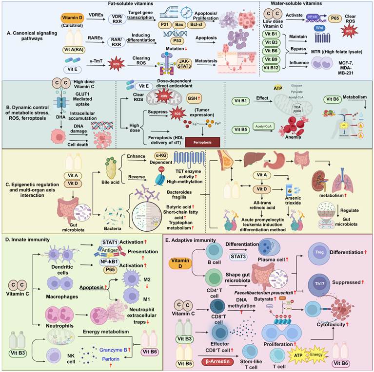
Vitamins affect cancer not merely through nutritional supplementation, but through coordinated regulation of tumor-intrinsic states, host metabolism, the tumor microenvironment (TME), including antitumor immunity. Fat-soluble vitamins often act through nuclear receptor signaling, membrane stability, antioxidation, and inflammatory regulation, whereas water-soluble vitamins mainly participate in coenzyme reactions, one-carbon metabolism, redox control, and epigenetic regulation. These processes ultimately affect proliferation, differentiation, death, metabolic adaptation, DNA damage responses, immune escape, and therapeutic sensitivity. The following section summarizes these preclinical mechanistic studies. Figure 3 summarizes the major mechanisms, and Table 2 lists representative vitamins, targets, and tumor-related implications.
Representative mechanisms and molecular targets of vitamins in cancer and their potential therapeutic implications
| Key vitamin(s) | Mechanistic category | Main targets/pathways | Tumor phenotypes and potential therapeutic implications | References |
|---|---|---|---|---|
| Vitamin C | TET-dependent DNA demethylation | TET2/TET3; 5hmC; stem-cell self-renewal and differentiation | Restores hydroxymethylation, limits aberrant hematopoietic self-renewal and leukemogenesis, and reduces malignant phenotypes in melanoma and bladder cancer models. TET restoration also increases leukemia-cell sensitivity to PARP inhibition. | [81-85] |
| Vitamin C | Histone remodeling and targeted-therapy sensitization | HDAC1-histone acetylation-BRD4; KDM5-H3K4 methylation-AKT2/mTOR | HDAC1 induction reduces histone acetylation and BET-inhibitor requirements; KDM5-dependent H3K4 demethylation enhances PI3K-inhibitor activity in triple-negative breast cancer models. | [72, 73] |
| Vitamin C | miRNA-mediated redox regulation | miR-93/miR-153-NRF2; antioxidant-response genes | Reduces estrogen-induced miR-93/miR-153 expression and restores NRF2-associated defenses in mammary carcinogenesis models. This context-specific preventive mechanism differs from pharmacological pro-oxidant killing. | [86-88] |
| Vitamin C | Pharmacological pro-oxidant cytotoxicity | Extracellular H2O2; oxidative injury; redox-dependent cell death | Pharmacological ascorbate generates extracellular H2O2 and selectively kills susceptible cancer cells; tumor inhibition is supported by cell and xenograft experiments. Delivery route and achievable exposure are critical. | [67, 89, 90] |
| Vitamin C | Glycolytic blockade | GLUT1-mediated DHA uptake; glutathione depletion; GAPDH oxidation and inhibition | In KRAS/BRAF-mutant colorectal cancer models, DHA reduction increases oxidative stress, inhibits GAPDH and depletes ATP. | [91] |
| Vitamin C | Hypoxia-response regulation | HIF-1α abundance/activity; HIF hydroxylases; GLUT1 and VEGF | Physiological ascorbate repletion moderates HIF signaling and tumor growth; tumor-tissue studies show inverse associations between ascorbate and HIF activity. Pharmacological ascorbate can also suppress HIF-1α through H2O2-dependent, hydroxylase-independent mechanisms. | [92-97] |
| Vitamin C | Transporter-dependent vulnerability and cell-cycle arrest | SVCT2; ROS; DNA damage; ATP depletion; p53/p21; Bax/Bcl-2 | SVCT2-dependent uptake sensitizes liver cancer stem cells to oxidative injury. Oral squamous carcinoma models show apoptosis, G0/G1 arrest and cisplatin sensitization. HIF activation increases ascorbate sensitivity in selected models, but is not a universal response predictor. | [98-100] |
| Vitamin C | Tumor antigen presentation and T-cell recruitment | IFN-γ-JAK/STAT-TET2; MHC-I machinery; CXCL9/10/11 | TET2-dependent demethylation increases tumor antigen presentation, chemokine expression and lymphocyte infiltration; ascorbate improves anti-PD-L1 responses in preclinical melanoma/colon tumor models. | [70, 101] |
| Vitamin C | Protein vitcylation and interferon signaling | STAT1 K298 vitcylation; reduced TCPTP binding; sustained STAT1 Y701 phosphorylation | Enhances interferon signaling and MHC/HLA-I expression, improving tumor recognition and antitumor T-cell responses in experimental models. | [102] |
| Vitamin C | Dendritic-cell differentiation and T-cell priming | NF-κB/p65-TET2-dependent demethylation; antigen-presentation genes; p38/ERK-IL-12p70 | Human monocyte-derived DCs acquire enhanced immunogenic properties, while murine DCs favor Th1 polarization. These are cellular immune mechanisms; the cited experiments do not establish clinical cancer-treatment efficacy. | [19, 69] |
| Vitamin C | Inflammatory signaling | TNF/IL-1-NIK/IKK-IκBα-NF-κB; context-dependent p38 signaling | Inhibits stimulus-induced NF-κB activation in cultured cell systems. The inhibitory effect is context dependent and should be distinguished from NF-κB-driven DC reprogramming. | [103, 104] |
| Vitamin C | Tumor-associated macrophage remodeling | Tumor-targeted vitamin C liposomes; M2-to-M1-like polarization; cytotoxic T-cell infiltration | Targeted vitamin C delivery remodels suppressive macrophages and improves anti-PD-L1 efficacy in mouse tumor models; effects depend on the tested delivery formulation. | [105] |
| Vitamin C and vitamin E | NET formation and redox balance | ROS; antioxidant capacity; GSH/GSSG; LPS-induced NET formation | Antioxidants reduce NET formation in isolated human neutrophils; combination effects involve vitamin C/E, glutathione and N-acetylcysteine. No tumor model was tested, so this supports a potential mechanism rather than demonstrated antitumor benefit. | [106] |
| Vitamin C | NK-cell expansion for adoptive therapy | Phosphorylated ascorbate; NK-cell proliferation and maturation | Increases ex vivo NK-cell yield while preserving tumor-cell killing under normoxia and hypoxia. The study supports cell-manufacturing optimization, rather than improved NK cytotoxicity per cell or established clinical efficacy. | [107] |
| Vitamin C | B-cell differentiation and immune homeostasis | TET2/3-Prdm1 demethylation; IL-21/STAT3; TET-associated Treg stability | Facilitates plasma-cell differentiation in primary immune-cell experiments. TET/vitamin C effects on Tregs are reviewed separately; immune homeostasis should not be equated with uniformly enhanced tumor rejection. | [108, 109] |
| Vitamin C | Vascular remodeling and immune accessibility | TET2-IL-2/STAT5A-tumor cGAS; cGAMP-LRRC8C-endothelial STING; VEGF/bFGF/MMP2 | Promotes vascular normalization and lymphocyte trafficking, enhancing anti-PD-L1 ± IL-2 in liver cancer models. Separate sarcoma experiments associate ascorbate with reduced angiogenic-gene expression and tumor growth. | [68, 110] |
| Vitamin C | Dose-dependent extracellular-matrix regulation | MMP-1/MMP-2; TGF-β; cancer-cell proliferation | An in vitro study reports biphasic effects on cancer-cell viability, MMPs and TGF-β. The direction of growth and matrix effects varies with concentration; an in vivo metastatic or vascular benefit was not demonstrated. | [111] |
| Vitamin C derivative | Bone metastatic niche remodeling | Ascorbate-2-phosphate magnesium; cell-cycle arrest; osteoclast differentiation; neutrophil recruitment/CXCR2 | Suppresses renal-cell-carcinoma stem-like phenotypes and bone metastasis; CXCR2 blockade further reduces progression in mice. The disease context is renal cancer bone metastasis. | [77] |
| Vitamin C | Radiotherapy-associated stromal injury | Neutrophil-derived S100A8/S100A9; fibroblast-to-myofibroblast differentiation | Attenuates radiation-induced pulmonary fibrosis while preserving or improving tumor control in mouse experiments. This concerns treatment-associated normal-tissue injury as well as therapeutic compatibility. | [64] |
| Vitamin D | Nuclear-receptor signaling and differentiation | 1,25(OH)2D-VDR/RXR; cell-cycle and differentiation programs; CYP27B1/CYP24A1; androgen signaling | Cancer-focused reviews describe context-dependent effects on proliferation, apoptosis and stromal signaling. New prostate organoid/cell/mouse experiments link deficiency to impaired luminal differentiation and show duration-dependent responses to calcitriol. | [112-116] |
| Vitamin D | RNA m6A regulation | VDR binding to the METTL14 promoter; m6A-related gene regulation | Vitamin D/VDR increases METTL14 and reduces tumor burden in colitis-associated colorectal cancer models, linking receptor signaling to epitranscriptomic regulation. | [117] |
| Vitamin D | Alternative splicing and histone regulation | 25(OH)D-KHDRBS3 downregulation; KDM6A Δexon13; H3K27me3-TRAP1-Smad2/3 | Promotes KDM6A exon-13 skipping, represses TRAP1 and reduces breast cancer proliferation/stemness; GSK-J4 combination shows preclinical synergy. | [118] |
| Vitamin D | Ferroptosis and cancer stemness | SLC7A11 downregulation; cysteine/GSH depletion; ROS and lipid injury | Induces ferroptotic phenotypes and reduces colorectal cancer stem-cell growth in cell and mouse models, suggesting a preclinical vulnerability of stem-like cells. | [119] |
| Vitamin D | Microbiota-barrier-systemic immunity | Intestinal epithelial VDR; Bacteroides fragilis; epithelial barrier integrity; microbial translocation | Intestinal VDR-dependent microbiota changes promote immune-mediated tumor control and checkpoint-blockade responses. Separate gut-breast experiments show that epithelial VDR loss causes dysbiosis/barrier failure and greater breast tumorigenesis. | [15, 16, 120, 121] |
| Vitamin D | Microbial vitamin metabolism | Estrogen-dependent C. maltaromaticum colonization; F. prausnitzii cross-feeding; 7-dehydrocholesterol; mucosal VDR | Microbial cooperation increases intestinal vitamin D-related metabolism and suppresses colorectal tumorigenesis in female mice. Sex dependence and microbial context limit generalization. | [122] |
| Vitamin D | Fibroblast-tumor communication | Calcitriol-treated CAFs; exosomal miR-378c; KDSR suppression | Reprograms CAF-derived exosomal cargo to inhibit gastric cancer migration, invasion and tumor growth. miR-378c experiments also show changes consistent with enhanced antitumor immunity. | [123] |
| Vitamin D | Oxidative stress and neutrophil recruitment | Prostate epithelial VDR/PTEN; oxidative stress; CXCL5-CXCR2 signaling | Epithelial VDR loss increases prostate cancer aggressiveness and neutrophil-associated dissemination. Blocking neutrophil chemotaxis reduces liver micrometastases in mice; this is not a vitamin D supplementation trial. | [124] |
| Vitamin D | Dendritic-cell and myeloid immune regulation | Monocyte-derived DC differentiation/maturation; costimulation; IL-1β/IL-6/TNF; TLR2/TLR4 | Calcitriol inhibits full DC differentiation and alters inflammatory mediators/chemotaxis in a maturation-dependent manner. These non-tumor immune-cell experiments do not establish uniform M2 polarization or tumor immunosuppression. | [125, 126] |
| Vitamin A and retinoids | Differentiation and growth control | RA-RAR/RXR; differentiation programs; p53-associated changes | Retinoic acid and tested metal complexes inhibit EJ bladder cancer growth and alter differentiation-associated phenotypes in vitro. Bladder cancer mechanisms are also summarized in a review; a microbiota-mediated clinical benefit is not established. | [35, 127] |
| Vitamin A and retinoids | Senescence remodeling and NK-cell surveillance | RAR/RXR agonists; p21; senescence-associated secretory phenotype; NK recruitment/activation | Retinoids, including adapalene, reprogram docetaxel-induced senescence and enhance NK-dependent tumor control in prostate cancer models. Pharmacological retinoids are distinct from dietary vitamin A. | [128] |
| Vitamin A and retinoids | Autocrine DC tolerance | ALDH1A2-derived retinoic acid; DC maturation and costimulation | DC-intrinsic retinoic acid restrains maturation. Genetic or pharmacological ALDH1A2 inhibition releases this brake and improves antitumor DC-vaccine efficacy in experimental models. | [129] |
| Vitamin A and retinoids | Radiotherapy-myeloid-T-cell cooperation | ATRA; IFN-γ; TNF/iNOS-producing inflammatory macrophages | ATRA plus radiotherapy induces inflammatory macrophages, increases effector T-cell recruitment and improves local/abscopal tumor control, including combinations with anti-PD-L1, in mice. | [65] |
| Vitamin B2 | FSP1-dependent ferroptosis resistance | Riboflavin-RFK-FMN-FAD; FSP1 cofactor binding/stability; RNF8-mediated proteasomal degradation | FAD stabilizes and supports FSP1 antioxidant recycling. Riboflavin-pathway disruption or the antimetabolite roseoflavin sensitizes susceptible cancer cells to ferroptosis; these are mechanistic preclinical findings. | [130, 131] |
| Vitamin B2 | Tumor ferroptosis resistance and immune exclusion | FAD synthase/FLAD1; FAD production; PCBP2 stabilization; cGAS-STING | FAD synthase supports hepatocellular carcinoma survival and restricts CD8+ T-cell recruitment. Genetic inhibition and hesperidin show antitumor activity in mice. | [132] |
| Vitamin B2 | Tumor-T-cell nutrient competition | Tumor SLC52A2 versus T-cell SLC52A3; mitochondrial respiration; GSH-GPX4; mitophagy and iron | Tumor riboflavin scavenging drives T-cell ferroptosis. Supplementation or engineered SLC52A3 expression improves CAR-T activity in preclinical models; the T-cell mechanism is FSP1 independent. | [133] |
| Vitamin B6 | Vitamin compartmentalization and tumor growth | Wnt-PRMT1/MrDegron; PDXK lysosomal microautophagy; intracellular PLP | Wnt-driven PDXK sequestration/degradation lowers active B6 and promotes colorectal cancer growth; disrupting this proteolytic pathway restores PLP and reduces growth in cell/organoid experiments. | [134] |
| Vitamin B6 | Tumor-NK-cell nutrient competition | Tumor one-carbon metabolism; NK-cell PLP-dependent glycogen breakdown | Pancreatic cancer cells consume B6 and impair NK-cell energy supply. B6 supplementation combined with tumor one-carbon-metabolism blockade restores NK activity and suppresses tumor burden in mice. | [9] |
| Vitamin B6 | CD8+ T-cell metabolic fitness and stemness | PLP-dependent transaminases; PDXK/PDXP; direct PLP-p70S6K binding; BACH2 nuclear retention | B6-dependent enzyme activity sustains T-cell proliferation/effector responses. A distinct PLP-p70S6K-BACH2 mechanism preserves stemness-like T cells and improves anti-PD-1 responses in preclinical models. | [135, 136] |
| Vitamin B5 | Transporter-dependent oncogenic metabolism | MYC-SLC5A6; pantothenate; coenzyme A; TCA-cycle/biosynthetic flux | MYC-high breast tumor regions accumulate pantothenate to support biosynthesis. Dietary restriction reverses metabolic adaptations and slows tumor growth in mice, indicating context-dependent tumor nutrient dependence. | [10] |
| Vitamin B5 | B-cell-T-cell cooperation and immunotherapy | Naïve B-cell-T-cell interaction; T-cell proliferation; IL-21/IFN-γ-associated responses | Human gastric cancer profiling/coculture experiments and mouse studies support B5-mediated immune cooperation; supplementation enhances anti-PD-1 responses in mouse models. Patient profiling is not proof of supplementation efficacy in patients. | [137] |
| Vitamin E and related tocols | DC antigen cross-presentation | SCARB1 uptake; direct SHP1 inhibition; MAPK; DC/extracellular-vesicle antigen presentation | Restores tumor-associated DC function and improves T-cell priming and checkpoint-blockade efficacy in mouse models. Patient survival associations are observational. | [17] |
| Vitamin B1 | Tumor microcirculation | Microvascular blood perfusion/flow velocity; neurogenic and endothelial oscillations; microvessel density | Thiamine inhibits liver tumor growth and improves microhemodynamic measures in mice. A molecular causal pathway and synergy with chemotherapy/immunotherapy were not established. | [138] |
| Vitamin B3 | Myeloid suppression and CD8+ T-cell activity | GPR109A-β-arrestin-mediated receptor degradation; NF-κB inhibition | Dietary B3 reduces suppressive myeloid polarization and restores CD8+ T-cell cytotoxicity, improving immunotherapy or targeted therapy in liver cancer models. | [71] |
| Vitamin B12 | Methionine self-sufficiency | B12-dependent methionine synthase/MTR; homocysteine remethylation; 4-ethylphenylcobalamin | B12 deprivation/antagonism sensitizes methionine-self-sufficient cancers to methionine restriction, including pancreatic xenografts. This is an experimental antagonism strategy, not evidence that B12 supplementation treats cancer. | [139] |
| Vitamin K and derivatives | Cell survival, senescence and redox regulation | K1/K2/K3-specific effects; p21/Bcl-2; mitochondrial redox signaling; FSP1-vitamin K hydroquinone | Review-level evidence links selected derivatives to growth arrest, apoptosis/autophagy and senescence-related signaling. Conversely, FSP1-dependent vitamin K recycling suppresses lipid peroxidation/ferroptosis. These opposing effects preclude a uniform anticancer conclusion. | [42] |
| Multiple B vitamins | Host metabolism and cancer cachexia | Hepatic niacin/B6/cofactor-associated enzymes; glycine-related one-carbon metabolism; protein malonylation | Mouse multi-omics and patient blood analyses identify reduced B-vitamin-related enzymes/metabolites during cachexia. Findings are associative and do not demonstrate that vitamin supplementation reverses wasting. | [140] |
Abbreviations: ATRA: all-trans retinoic acid; BET: bromodomain and extra-terminal; CAF: cancer-associated fibroblast; CAR-T: chimeric antigen receptor T cell; CoA: coenzyme A; DC: dendritic cell; DHA: dehydroascorbic acid; ECM: extracellular matrix; FAD: flavin adenine dinucleotide; FMN: flavin mononucleotide; FSP1: ferroptosis suppressor protein 1; GSH/GSSG: reduced/oxidized glutathione; HIF: hypoxia-inducible factor; IFN: interferon; MHC: major histocompatibility complex; MMP: matrix metalloproteinase; MTR: methionine synthase; NET: neutrophil extracellular trap; NK: natural killer; PLP: pyridoxal 5′-phosphate; RA: retinoic acid; RAR/RXR: retinoic acid receptor/retinoid X receptor; ROS: reactive oxygen species; SAM: S-adenosylmethionine; SVCT2: sodium-dependent vitamin C transporter 2; TCA: tricarboxylic acid; TCPTP: T-cell protein tyrosine phosphatase; TET: ten-eleven translocation; THF: tetrahydrofolate; TME: tumor microenvironment; VDR: vitamin D receptor; 5hmC: 5-hydroxymethylcytosine.
Vitamin-mediated molecular regulation in cancer signaling, metabolism, and immunity. (A) Signaling networks: Fat-soluble vitamins (D, A, E) regulate transcription, apoptosis, and metastasis via nuclear receptors/antioxidant pathways; water-soluble vitamins (C, B-complex) modulate pro-survival and metabolic pathways. (B) Metabolism and ferroptosis: High-dose vitamin C induces tumor cell death; vitamin E regulates ferroptosis via antioxidant/iron-dependent ROS modulation; B-vitamins support mitochondrial energy production. (C) Epigenetics and gut-axis crosstalk: Vitamins A/D modulate the gut microbiota-organ axis, regulate TET-mediated epigenetic reprogramming, and support systemic homeostasis and leukemia differentiation. (D) Innate immunity: Vitamins C/B enhance dendritic cell antigen presentation, macrophage polarization, and NK cell cytotoxicity. (E) Adaptive immunity: Vitamin D regulates T/B cell differentiation; vitamins C/B3/B5 boost CD8⁺ T cell anti-tumor function via epigenetic and signaling modulation. Abbreviations. γ-TMT: γ-Tocopherol Methyltransferase; Vit A: vitamin A; Vit B1: thiamine; Vit B3: niacin; Vit B5: pantothenic acid; Vit B6: pyridoxine; Vit B9: folate; Vit B12: cobalamin; Vit D: vitamin D; Vit E: vitamin E; RA: retinoic acid; VDREs: vitamin D response elements; VDR: vitamin D receptor; RXR: retinoid X receptor; Bax: BCL2-associated X protein; Bcl-xL: B-cell lymphoma-extra large; RAREs: retinoic acid response elements; RAR: retinoic acid receptor; ROS: reactive oxygen species; JAK: Janus kinase; STAT3: signal transducer and activator of transcription 3; NF-κB: nuclear factor kappa B; p65: NF-κB p65 subunit (RelA); MTR: methionine synthase; GLUT1: glucose transporter 1; DHA: dehydroascorbic acid; GSH: glutathione; HDL: high-density lipoprotein; CoA: coenzyme A; TCA: tricarboxylic acid; α-KG: alpha-ketoglutarate; TET: ten-eleven translocation dioxygenase; STAT1: signal transducer and activator of transcription 1; M1/M2: M1-like/M2-like macrophages; NK: natural killer; Treg: regulatory T cell; Th17: T helper 17 cell; SAM: S-adenosylmethionine; SAH: S-adenosylhomocysteine.
Effects on tumor cells
Nuclear receptor-mediated transcriptional regulation
Nuclear receptor signaling is a classic mechanism by which fat-soluble vitamins regulate tumor cells. The active form of vitamin D, 1,25(OH)₂D, binds the vitamin D receptor (VDR), forms a heterodimer with retinoid X receptor (RXR), and binds vitamin D response elements to regulate genes involved in cell-cycle control, differentiation, adhesion, metabolism, and epithelial-mesenchymal transition [112, 141, 142]. VDR signaling also crosstalks with Wnt/β-catenin, MAPK/ERK, PI3K/Akt, and p53 pathways, thereby affecting proliferation, migration, apoptosis, and therapy response [113-115]. Thus, vitamin D activity depends not only on circulating levels, but also on tumor VDR expression, nuclear localization, and vitamin D metabolic enzymes. Upregulation of degradative enzymes such as CYP24A1 may weaken active vitamin D signaling and promote vitamin D resistance [115, 121].
Vitamin A mainly acts through its active metabolite retinoic acid (RA). RA binds RA receptors (RARs), forms complexes with RXR, and regulates genes involved in differentiation, apoptosis, senescence, and tissue organization through RA response elements [35, 143]. In many tumors, RA suppresses proliferation, induces differentiation, and promotes apoptosis. The success of ATRA in acute promyelocytic leukemia further shows that vitamin derivatives can be translated into standard anticancer therapy by relieving differentiation arrest [76]. However, RA/RAR/RXR signaling is tissue-specific and must be interpreted according to cancer type, differentiation state, and immune context.
Epigenetic remodeling
Epigenetic dysregulation is central to tumorigenesis, and some vitamins act as cofactors or substrates for epigenetic enzymes. Vitamin C is a representative epigenetic regulator. TET enzymes require Fe²⁺ and α-ketoglutarate and convert 5-methylcytosine to 5-hydroxymethylcytosine, mediating active DNA demethylation [85]. Vitamin C enhances TET activity, promotes demethylation of aberrantly hypermethylated regions, and restores tumor-suppressive and differentiation-related genes [81, 82]. In acute myeloid leukemia, melanoma, and bladder cancer models, vitamin C suppresses malignant progression by increasing 5hmC [82-84], and also regulates histone demethylases and miRNA networks [86-88].
B vitamins contribute to epigenetic regulation through distinct but interconnected one-carbon and amino acid metabolic pathways. Folate supplies one-carbon units for nucleotide synthesis and homocysteine remethylation, while vitamin B12 serves as a cofactor for methionine synthase, supporting methionine regeneration and subsequent S-adenosylmethionine (SAM) synthesis. Vitamin B6, primarily in its active pyridoxal 5′-phosphate (PLP) form, functions as a cofactor for serine hydroxymethyltransferase and transsulfuration enzymes; it thereby influences one-carbon-unit availability and sulfur amino acid metabolism rather than directly catalyzing homocysteine remethylation. These pathways can help maintain normal methylation and genomic stability, whereas their dysregulation may support nucleotide synthesis and methylation demands in established tumors [13, 144]. Vitamin D may also regulate RNA epigenetics. VDR was recently shown to regulate the m6A methyltransferase METTL14 in colitis-associated CRC; vitamin D treatment increased METTL14 and reduced tumor burden, whereas Vdr deletion decreased METTL14 and promoted tumorigenesis [117]. This extends vitamin D biology from transcriptional regulation to m6A RNA modification.
Metabolic reprogramming
Vitamins can also directly affect energy metabolism, biosynthesis, and stress adaptation. Vitamin C mainly regulates glycolysis and hypoxic adaptation. In KRAS- or BRAF-mutant colorectal cancer cells, increased GLUT1-mediated uptake of dehydroascorbate can deplete intracellular glutathione during its reduction to ascorbate, leading to oxidative stress and GAPDH inactivation. This impairs glycolytic flux and causes an energetic crisis in these highly glycolysis-dependent cells [91]. Separately, ascorbate supports HIF hydroxylase activity and can attenuate HIF-1 signaling in experimental cancer models [96, 145]. An inverse association between tumor ascorbate levels and hypoxia-pathway activation has also been reported in human breast cancer tissue [92]. B vitamins participate in broader metabolic networks. Vitamin B5 accumulates in MYC-high breast tumor regions and is converted into coenzyme A to support TCA-cycle activity and biosynthesis. MYC upregulates the multivitamin transporter SLC5A6 to increase B5 uptake, whereas dietary B5 restriction reverses MYC-driven metabolic changes and suppresses tumor growth [10]. Thus, vitamins are not passive nutrients, but components of spatial metabolic heterogeneity and tumor adaptation.
Redox homeostasis and cell death
Redox regulation is a key interface between vitamins and cancer. Vitamin E is a lipid-soluble antioxidant that scavenges lipid peroxyl radicals and reduces membrane lipid peroxidation [53, 146]. Vitamin C acts mainly as an antioxidant and enzymatic cofactor at physiological concentrations, but at pharmacological concentrations, especially after intravenous delivery, it can become a pro-oxidant that damages tumor cells through hydrogen peroxide generation, iron metabolism disturbance, and ROS accumulation [67, 77, 98, 100, 147]. This dose-dependent antioxidant-to-pro-oxidant switch is central to its anticancer activity.
Vitamin-induced redox changes could also affect DNA damage responses and finally induce cell death. Pharmacological vitamin C enhances DNA damage, cell-cycle arrest, and apoptosis, and synergizes with DNA damage-dependent therapies such as radiotherapy and chemotherapy [64, 67, 89-91, 110]. Vitamin D upregulates pro-apoptotic proteins such as Bax and Bad and suppresses anti-apoptotic signaling [114, 115]. Recent work also links vitamin D to ferroptosis: in CRC stem-like cells, vitamin D downregulates SLC7A11, lowers cysteine and glutathione, increases ROS, and induces ferroptosis; SLC7A11 overexpression attenuates this effect [119]. In hepatocellular carcinoma, vitamin B2-related FAD synthase confers ferroptosis resistance and limits CD8⁺ T-cell recruitment, linking flavin metabolism to cell death resistance and immune escape [132]. These observations place vitamin-regulated redox networks within a broader ferroptosis framework in which altered lipid-peroxidation sensitivity can contribute to both therapeutic resistance and metastatic competence. Rational vitamin-based combinations should therefore consider the GPX4/GSH axis, iron handling, and context-dependent immune effects on ferroptosis susceptibility [148].
Inflammatory and oncogenic signaling
Chronic inflammation promotes tumorigenesis and alters treatment response. Beyond antioxidation, vitamin E inhibits COX, 5-LOX, NF-κB, JAK/STAT6, and JAK/STAT3 signaling [146, 149]. In cellular models, vitamin C inhibits TNF-α-induced NF-κB activation by reducing IκBα phosphorylation [103]. A separate study linked vitamin C-mediated inhibition of TNF-induced NF-κB activation to p38 MAPK activation in endothelial cells [104]. Vitamin D also regulates inflammation through VDR signaling and interacts with PI3K/Akt/mTOR, MAPK, and p53 pathways [113-115]. Together, these effects provide a basis for suppressing pro-inflammatory, pro-survival, and resistance-related signaling.
Effects on the TME
Gut microbiota and mucosal barrier
The tumor immune microenvironment (TIME) is shaped not only by local tumor factors, but also by gut microbiota, mucosal barrier integrity, and systemic immunity. Vitamin D is representative in this regard. VDR in intestinal epithelial cells maintains tight junctions and barrier integrity by upregulating occludin, E-cadherin, and ZO-1, thereby limiting dysbiosis and systemic exposure to harmful metabolites [113, 120]. Vitamin D also reshapes the gut microbiota and enhances CD8⁺ T-cell activation through the MyD88-IFN-I-cDC1 axis [15, 150]. Recent studies further reveal bidirectional links between microbiota and vitamin D metabolism. Carnobacterium maltaromaticum, depleted in female CRC patients, cooperates metabolically with Faecalibacterium prausnitzii to convert 7-dehydrocholesterol into vitamin D-related metabolites, activate mucosal VDR signaling, and suppress CRC [122]. Intestinal epithelial VDR loss can also drive dysbiosis, barrier disruption, microbial translocation, and inflammation, thereby promoting extraintestinal breast tumorigenesis [16]. Thus, vitamin D may act through a microbiota-barrier-VDR-systemic immunity axis.
Vitamin A also contributes to intestinal mucosal immune regulation through its metabolite RA. RA induces the gut-homing receptors α4β7 and CCR9 on activated T cells, thereby promoting their migration to the intestine [151]. In addition, RA cooperates with TGF-β to support the induction of Foxp3⁺ regulatory T cells by intestinal CD103⁺ dendritic cells, providing a mechanism for the regulation of mucosal immune tolerance [152].
Innate immunity, antigen presentation, and myeloid remodeling
Innate immune cells are key components of the TIME. Vitamin D usually acts as an immune homeostasis regulator. It suppresses monocyte differentiation into mature dendritic cells (DCs), reduces MHC-II and costimulatory molecule expression, increases IL-10, and decreases IL-12 [121, 125]. In human monocytes and macrophages, 1,25-dihydroxyvitamin D3 modulates inflammatory mediator and Toll-like receptor expression in a maturation-dependent manner and also affects macrophage chemotaxis [126]. These findings indicate that vitamin D regulates innate immune function in a cell state-dependent manner; its consequences for antitumor immunity require evaluation in specific tumor contexts.
Vitamin E, B3, and C are more closely associated with antitumor immune priming or relief of myeloid suppression. Vitamin E enters DCs through SCARB1, inhibits SHP1, activates MAPK signaling, and enhances antigen presentation and T-cell priming [17]. Vitamin B3 promotes GPR109A degradation through GPR109A/β-arrestin signaling, inhibits NF-κB activation, reverses myeloid immunosuppression, and restores CD8⁺ T-cell cytotoxicity [71]. Vitamin C-loaded liposomes promote the repolarization of tumor-associated macrophages from an M2-like toward an M1-like phenotype [105]. Separately, vitamin C reduces lipopolysaccharide (LPS)-induced neutrophil extracellular trap formation in isolated human neutrophils in vitro [106]. It also enhances tumor antigen presentation and T-cell IFN-γ responses through TET2 [101]. A newer mechanism is vitamin C-derived lysine vitcylation: STAT1 K298 vitcylation prevents STAT1 interaction with the phosphatase TCPTP, enhances STAT1-mediated interferon responses and MHC/HLA-I expression, and promotes antigen presentation and T-cell antitumor immunity [102].
The seemingly contradictory immunomodulatory effects of vitamin D should be understood as context-dependent rather than mutually exclusive. At physiological or repletion doses, VDR signaling in intestinal epithelial cells and cDC1s may maintain barrier integrity and support microbiota-dependent MyD88-IFN-I-cDC1-CD8+ T cell antitumor immunity. [15, 150]; at pharmacological or supraphysiological exposure, however, it may promote tolerogenic dendritic cells, M2-like macrophages, and Th2/Treg-biased responses, while suppressing antigen presentation [153-155]. Tissue location is equally important, because intestinal VDR signaling, tumor-intrinsic VDR/CYP24A1 activity, and myeloid/lymphoid cell populations in the tumor microenvironment or draining lymph nodes may respond differently to the same systemic 25(OH)D level. Therefore, baseline vitamin D status and local immune cell composition together determine whether the predominant effect is immune activation or immune suppression. Dose, route, timing, tissue site, tumor stage, baseline 25(OH)D, and the local immune context should be integrated to predict whether vitamin D promotes or restrains antitumor immunity.
Adaptive immunity
Adaptive immunity, especially CD8⁺ T-cell cytotoxicity, is central to antitumor immunotherapy. Vitamin C can also enhance antitumor immunity through T-cell-dependent mechanisms. Using lymphocyte depletion and adoptive-transfer experiments, Magrì et al. showed that CD8⁺ T cells mediate much of its antitumor activity, with CD4⁺ T cells contributing to the acquisition of effective CD8⁺ T-cell cytotoxicity [18]. Vitamin C also enhances TET2-dependent expression of IFN-γ-responsive chemokines in tumor cells, promotes lymphocyte infiltration, and improves the efficacy of anti-PD-L1 therapy in preclinical tumor models [70]. It also activates TET2 in tumor cells, upregulates cGAS, promotes cGAMP production, and activates STING signaling through LRRC8C-mediated cGAMP release, thereby inducing vascular normalization and lymphocyte infiltration [68]. Vitamin B3 increases Ki67, perforin, granzyme B, IFN-γ, and TNF-α in tumor-infiltrating CD8⁺ T cells, enhancing proliferation and cytotoxicity [71]. Vitamin B6 provides a distinct mechanism related to T-cell persistence: its active form pyridoxal phosphate preserves stemness-like features and antitumor ability of tumor-infiltrating CD8⁺ T cells [136], and B6 metabolism determines T-cell antitumor responses [135]
For NK cells, PDAC cells consume large amounts of vitamin B6 to support one-carbon metabolism and growth, thereby limiting B6 availability for NK cells, which need it for glycogen breakdown and activation. Vitamin B6 supplementation combined with one-carbon metabolism blockade restores NK-cell antitumor function and reduces tumor burden [9]. These findings show that vitamins are functional resources competed for by tumor and immune cells. By contrast, vitamin D preferentially regulates immune balance by suppressing Th1 and Th17 differentiation and promoting Th2 and anti-inflammatory responses [153-155]. Vitamin C can also stabilize Foxp3 expression in Tregs through TET-mediated demethylation and promote B-cell differentiation into plasma cells [108, 109].
Other components in the TME
Tumor vasculature, stromal structure, and metastatic niches determine immune-cell entry and drug accessibility. Vitamin C induces vascular normalization and lymphocyte infiltration through the TET2-cGAS-STING axis, linking epigenetics, innate immune sensing, and adaptive immune amplification [68]. A stable vitamin C derivative suppresses renal cancer bone metastasis by inducing cell-cycle arrest, reducing stem-like features, and remodeling the bone metastatic microenvironment through inhibition of osteoclast differentiation and neutrophil recruitment; combination with a CXCR2 antagonist further suppresses bone metastasis [77]. Systemic host metabolism is also relevant. In cancer cachexia, reduced liver enzymes related to niacin, vitamin B6, and glycine-linked one-carbon metabolism represent a systemic metabolic feature and are reflected in blood samples from gastric cancer patients with cachexia [140].
Safety, contraindications, and drug interactions
Much of the existing evidence is derived from short-term studies or oncologic cohorts with limited sample sizes, making it impossible to draw definitive conclusions regarding the safety of vitamin supplementation. When administering vitamins, their safety should be individually assessed according to each cancer patient's specific condition. Some studies have also demonstrated that vitamins may reduce therapeutic efficacy when used in combination with certain treatment modalities. For instance, coadministration of vitamin C reduced imatinib bioavailability in rats [156]. In preclinical multiple myeloma models, vitamin C also attenuated the antitumor activity of bortezomib; the observed antagonism was restricted to boronate-based proteasome inhibitors, indicating that the interaction depends on drug-specific properties [157].
Ongoing vitamin-related clinical trials in oncology
The studies discussed above suggest that vitamins may influence cancer biology by regulating tumor metabolism, epigenetic status, oxidative stress, the immune microenvironment, and treatment sensitivity. These mechanistic insights have prompted increasing clinical trials to evaluate the translational value of vitamin-based interventions in cancer prevention and treatment. Current trials mainly focus on four areas. First, vitamins are used to correct deficiency and improve supportive care outcomes, with particular attention to vitamin D in pain, infection risk, bone metabolism, muscle function, quality of life, and endocrine therapy-related bone loss in patients with advanced cancer. Second, vitamins are being tested as treatment sensitizers, especially high-dose intravenous vitamin C combined with chemotherapy, radiotherapy, or targeted therapy in pancreatic, gastric, liver, and other solid tumors. Third, studies are assessing whether baseline vitamin status affects responses to chemoradiotherapy, immunotherapy, or multimodal treatment, thereby informing patient stratification. Fourth, vitamins or their derivatives are being explored in high-risk populations, premalignant lesions, postoperative recurrence prevention, and long-term cancer management. Overall, most ongoing trials remain early-stage, exploratory, or focused on supportive endpoints, with limited sample sizes and stratified designs. Future studies should integrate baseline vitamin levels, cancer type, molecular subtype, immune phenotype, and treatment regimen to identify patients most likely to benefit from vitamin interventions. Representative trials are summarized in Table 3.
Registered studies of vitamins and vitamin derivatives in cancer treatment, prevention, and supportive care
| Types of vitamins | NCT number | Recruitment status | Cancer type / setting | Vitamin formulation, dose, and route | Control group | Primary endpoint (s) |
|---|---|---|---|---|---|---|
| Vitamin D | NCT02172651 | RecruitingLast posted: 2026-05-13 | Stage I-III colon cancer or colon cancer with resectable liver metastases; preoperative intervention. | Cholecalciferol (D3), 50,000 IU orally once daily for 7 days, then 10,000 IU once daily until surgery (within 28 days). | Matching oral placebo in the randomized comparison; an initial vitamin D run-in cohort was also included. | VDR binding sites in malignant and normal colonic tissue (14-28 days); liver-metastasis TME changes in the hepatic-resection cohort. |
| Vitamin D | NCT05846880 | RecruitingLast posted: 2025-04-08 | Multiple myeloma undergoing ASCT and subsequent lenalidomide maintenance. | Cholecalciferol (D3), 50,000 IU weekly to correct deficiency until 25(OH)D >30 ng/mL, then 50,000 IU monthly as maintenance; route NR in the intervention description. | The same initial weekly vitamin D deficiency correction, followed by no maintenance vitamin D; lenalidomide in both groups. | Absolute lymphocyte count and CD4/CD8 lymphocyte subsets at 120 days after ASCT. |
| Vitamin D | NCT06596122 | Not yet recruitingLast posted: 2024-09-19 | Breast cancer receiving AC-T chemotherapy; prevention of paclitaxel-induced peripheral neuropathy. | Cholecalciferol (D3), 50,000 IU orally weekly for 8 weeks, then 100,000 IU monthly until chemotherapy completion, for deficient participants in the intervention group. | Standard AC-T chemotherapy alone. | Cumulative incidence and grade of paclitaxel-induced peripheral neuropathy during the 12-week paclitaxel phase (CTCAE v5.0). |
| Vitamin D | NCT04677816 | RecruitingLast posted: 2026-08-28 | Vitamin D-deficient invasive breast cancer receiving neoadjuvant chemotherapy. | Cholecalciferol (D3), 50,000 IU orally once weekly for 20 weeks from chemotherapy initiation. | Nonrandomized observational comparison with patients with normal vitamin D levels receiving standard neoadjuvant chemotherapy. | Number/proportion with pCR in the vitamin D-deficient supplementation group (up to 22 weeks). |
| Vitamin D | NCT06757244 | RecruitingLast posted: 2025-03-18 | Previously untreated metastatic pancreatic adenocarcinoma. | Cholecalciferol (D3), 8,000 IU orally daily for 14 days, then 4,000 IU daily, with dostarlimab and mFOLFIRINOX, followed by maintenance treatment. | None; single-arm combination study. | ORR by RECIST 1.1 (24 months). |
| Vitamin D | NCT05838716 | RecruitingLast posted: 2026-01-15 | Prostate cancer receiving ADT; prevention of treatment-associated bone loss. | Vitamin D, 50,000 IU orally once weekly for 52 weeks. Both groups also receive vitamin D 800 IU/day and calcium 1,000 mg/day. | Matching oral placebo once weekly for 52 weeks, with the same background vitamin D and calcium supplementation. | BMD loss at the total hip, femoral neck, distal radius, and lumbar spine at 52 weeks. |
| Vitamin D | NCT02779465 | Not yet recruitingLast posted: 2016-05-20 | Chronic hepatitis B on long-term antiviral therapy; HCC prevention. | Cholecalciferol (D3), 800 IU orally daily, added to nucleos(t)ide antiviral therapy. | Antiviral therapy without study vitamin D3 supplementation. | Change from baseline in serum 25(OH)D at 6 and 12 months. |
| Vitamin D | NCT07241585 | RecruitingLast posted: 2026-08-18 | Basal-cell or squamous-cell nonmelanoma skin cancer before Mohs surgery or ED&C. | Vitamin D (specific form NR), 10,000 IU orally daily for 6 days before PDT; surgery 1-14 days after PDT. | Oral placebo for 6 days plus PDT; an additional tissue-only group undergoes surgery without study supplementation or PDT. | Expression of immune-checkpoint molecules in tumors and peritumoral stroma at surgery (up to day 20). |
| Vitamin D | NCT05887505 | Not yet recruitingLast posted: 2023-06-06 | HCC undergoing hepatectomy; perioperative supplementation. | Cholecalciferol (D3), 576,000 IU orally daily for 3 consecutive days (total 1,728,000 IU), starting 7 days before surgery. | Oral placebo for 3 consecutive days. | Association between 25(OH)D and PTH; liver-failure proportion at registry-specified perioperative assessments. |
| Vitamin D | NCT06642441 | RecruitingLast posted: 2025-01-17 | Breast cancer receiving adjuvant chemotherapy. | Vitamin D2 (ergocalciferol), 10 mg by injection before chemotherapy and every 3 weeks until chemotherapy completion; injection route subtype NR. | Standard adjuvant chemotherapy alone. | Incidence and proportion of treatment-related adverse events during chemotherapy (4-6 months). |
| Vitamin C | NCT07121036 | RecruitingLast posted: 2025-08-13 | Advanced malignant solid tumors receiving standard systemic anticancer therapy. | Vitamin C, IV: 0.5 g/kg once daily, 0.5 g/kg twice daily, or 0.75 g/kg twice daily, for 5-7 days during the first systemic-treatment cycle. | No vitamin C-free control; three dosing cohorts, all receiving standard systemic therapy. | Vitamin C pharmacokinetics and blood-concentration changes from baseline to day 7. |
| Vitamin C | NCT06336902 | RecruitingLast posted: 2026-03-17 | Previously treated KRAS-mutant, MSS metastatic colorectal cancer. | Vitamin C, IV over 30 min on days 1, 15, and 29 of each 42-day cycle, with botensilimab, balstilimab, and FMD; numerical vitamin C dose NR. | None; single-arm combination study. | FMD adherence (≥75% of designated days) with all study-drug doses for ≥2 cycles; incidence of treatment-related adverse events (up to 30 months). |
| Vitamin C | NCT03999723 | RecruitingLast posted: 2024-04-17 | Higher-risk MDS, CMML-2, or low-blast-count AML receiving azacitidine. | Ascorbic acid, 1,000 mg orally once daily from day 1 of the first azacitidine cycle until azacitidine discontinuation or study end. | Matching oral placebo with azacitidine. | Event-free survival (up to 54 months). |
| Vitamin C | NCT06421688 | Unknown; last known: not yet recruitingLast posted: 2024-05-20 | Intracranial tumor surgery; prevention of postoperative pulmonary complications. | Ascorbic acid, 50 mg/kg IV (maximum 4 g), diluted to 50 mL and infused at 25 mL/h after anesthesia induction. | Saline, 50 mL IV at 25 mL/h after anesthesia induction. | Incidence of postoperative pulmonary complications during hospitalization (approximately 10 days). |
| Vitamin C | NCT06493370 | RecruitingLast posted: 2025-02-20 | Muscle-invasive bladder cancer in patients ineligible for or declining cisplatin-based neoadjuvant chemotherapy. | Ascorbic acid, initial 25-g IV dose, titrated to a target peak plasma concentration (numerical target NR), then twice weekly; combined with 2 cycles of gemcitabine/carboplatin. | None; single-arm study. | Post-treatment pathological stage/downstaging rate (approximately 10-12 weeks). |
| Vitamin C | NCT06018896 | Unknown; last known: recruitingLast posted: 2024-06-06 | Metastatic pancreatic adenocarcinoma progressing after two chemotherapy lines; quality-of-life intervention. | If baseline vitamin C <65 μmol/L: IV 10 g/day for 7 days, then 4 g/day for 7 days, then 2 g/day for 7 days, followed by oral 900 mg/day in 3 doses. If ≥65 μmol/L: oral 900 mg/day in 3 doses. | None; single-arm study. | Change in quality of life by EORTC QLQ-C30 at the end of cycle 1 (28 days). |
| Vitamin C | NCT06601283 | RecruitingLast posted: 2024-11-26 | Metastatic pancreatic adenocarcinoma receiving gemcitabine/nab-paclitaxel. | Vitamin C, 300 mg/day orally in 3 divided doses, with reishi and cordyceps (each 2 g/day orally in 2 doses) and gemcitabine/nab-paclitaxel. | None; single-arm study. | Rate of anemia at the end of cycle 1 (28 days). |
| Vitamin C | NCT04516681 | RecruitingLast posted: 2024-10-24 | Metastatic colorectal cancer with high GLUT3 expression. | Ascorbic acid, 1.5 g/kg/day IV on days 1-3 every 2 weeks, with adebrelimab and FOLFOX ± bevacizumab. | FOLFOX ± bevacizumab alone; adebrelimab is listed only in the experimental arm. | ORR (up to 5 years). |
| Vitamin B | NCT07257497 | Active, not recruitingLast posted: 2026-09-02 | Solid tumors continuing immunotherapy; dietary vitamin B6 restriction. | Vitamin B6-restricted liquid/food diet for 6 weeks (2 × 3-week cycles), targeting 30 kcal/kg/day and protein 1.5 g/kg/day; quantitative B6 intake limit NR. | None; single-arm diet-plus-immunotherapy study. | Type, frequency, and severity of treatment-emergent adverse events over 6 weeks. |
| Vitamin B | NCT07119996 | RecruitingLast posted: 2026-06-30 | Antibiotic-exposed cT2-T4aN0M0 bladder urothelial carcinoma; neoadjuvant chemotherapy plus tislelizumab. | Nicotinic acid (B3) tablets, 300 or 500 mg orally once daily after antibiotic treatment, with tislelizumab and gemcitabine/cisplatin. | None; single-arm dose-finding study. | Vitamin B3 dose-limiting toxicity during days 1-28. |
| Vitamin E | NCT05771181 | RecruitingLast posted: 2025-06-12 | MSS metastatic colorectal cancer. | Vitamin E (specific form NR), 400 mg orally once daily, with fruquintinib and tislelizumab, until progression or intolerable toxicity. | None; single-arm combination study. | ORR (2 years). |
| Vitamin A | NCT06371274 | RecruitingLast posted: 2025-08-12 | Unresectable locally advanced, recurrent, or metastatic triple-negative breast cancer. | ATRA, 150 mg/m²/day orally in 2 divided doses on days 0-2 of each 3-week cycle, with toripalimab, for up to 2 years. | None; single-arm combination study. | ORR by RECIST 1.1 (up to 2 years). |
| Vitamins D and K | NCT07256769 | RecruitingLast posted: 2025-12-01 | Breast cancer receiving endocrine therapy and initiating alendronate for bone-loss prevention. | Synostea®: calcium carbonate 400 mg, cholecalciferol 2,000 IU, vitamin K (menaquinone) 50 μg, α-lactalbumin 30 mg, and D-chiro-inositol 150 mg; route and frequency NR. | Standard supplementation with calcium carbonate 500 mg and cholecalciferol 2,000 IU; route and frequency NR. | Between-group difference in mean vitamin D levels at 6 months. |
| Vitamins B and C | NCT04150042 | RecruitingLast posted: 2026-01-29 | Metastatic pancreatic or breast cancer; chemotherapy with autologous hematopoietic stem-cell rescue (SHARON). | Hydroxocobalamin (B12b), 1.45 g/m² IV, plus vitamin C 3, 6, or 8 g/m² IV on day -2, with melphalan/BCNU; stem-cell infusion on day 0; 2 cycles approximately 6 weeks apart. Doses are from the 2023 trial report. | None; single-arm dose-escalation study. | Safety: sinusoidal obstruction syndrome, pulmonary toxicity, oxalate nephropathy, cytokine-release syndrome, mucositis, delayed/failed engraftment, and overall/serious/grade 3-5 adverse events (up to 12 months after cycle 2). |
| Vitamins A and B | NCT05702398 | RecruitingLast posted: 2026-03-20 | Skin-cancer prevention in high-risk kidney-transplant recipients with a history of skin cancer. | Retinyl palmitate (A), 1,000 μg orally twice daily, plus nicotinamide (B3), 500 mg orally twice daily, for 6 months. | Matching placebo pills twice daily. | Change in blood vitamin A and nicotinamide levels at 6 months. |
Abbreviations. 25(OH)D: 25-hydroxyvitamin D; AC-T: doxorubicin/cyclophosphamide followed by paclitaxel; ADT: androgen-deprivation therapy; AML: acute myeloid leukemia; ASCT: autologous stem-cell transplantation; ATRA: all-trans retinoic acid; BCNU: carmustine; BMD: bone mineral density; CMML-2: chronic myelomonocytic leukemia type 2; CTCAE: Common Terminology Criteria for Adverse Events; ED&C: electrodesiccation and curettage; EORTC QLQ-C30: European Organisation for Research and Treatment of Cancer Quality of Life Questionnaire Core 30; FMD: fasting-mimicking diet; FOLFOX: fluorouracil/leucovorin/oxaliplatin; FOLFOXIRI: fluorouracil/leucovorin/oxaliplatin/irinotecan; GLUT3: glucose transporter 3; HCC: hepatocellular carcinoma; IU: international units; IV: intravenous; MDS: myelodysplastic syndromes; mFOLFIRINOX: modified fluorouracil/leucovorin/irinotecan/oxaliplatin; MSS: microsatellite-stable; NCI: National Cancer Institute; NR: not reported; ORR: objective response rate; pCR: pathological complete response; PDT: photodynamic therapy; PTH: parathyroid hormone; RECIST: Response Evaluation Criteria in Solid Tumors; TME: tumor microenvironment; VDR: vitamin D receptor.
Conclusions and future perspectives
The roles of vitamins in cancer are complex and highly context-dependent. Current evidence suggests that adequate vitamin status may help maintain tissue homeostasis, reduce the risk of certain cancers, improve survival, or enhance treatment tolerance. However, vitamins should not be viewed simply as “anticancer factors.” High-dose or inappropriate supplementation may be ineffective or even harmful in specific cancer types, disease stages, or treatment settings. Therefore, vitamin-based interventions should move beyond empirical supplementation toward mechanism-guided and patient-stratified precision strategies.
Future studies should address several key issues. First, high-quality prospective cohorts and randomized trials are needed to determine whether vitamin status is a causal factor in cancer development or merely a marker of tumor burden, inflammation, or nutritional status. Second, the optimal dose, route, and safety boundary of vitamin interventions should be defined for different cancer types, molecular subtypes, and treatment stages. Third, predictive markers based on serum vitamin levels, metabolomics, gut microbiota, epigenetic status, and immune phenotypes should be developed for clinical stratification. Finally, interactions between vitamins and radiotherapy, chemotherapy, immunotherapy, or targeted therapy must be systematically assessed to avoid potential interference and maximize synergy. Only with clear mechanisms, rational dosing, and precise patient selection can the clinical value of vitamins in cancer prevention and treatment be fully realized.
Abbreviations
1,25(OH)₂D: 1,25-dihydroxyvitamin D; 25(OH)D: 25-hydroxyvitamin D; 5-LOX: 5-lipoxygenase; 5hmC: 5-hydroxymethylcytosine; AC-T: doxorubicin/cyclophosphamide followed by paclitaxel; ADT: androgen deprivation therapy; Akt: protein kinase B; ALDH1A2: aldehyde dehydrogenase 1 family member A2; α-KG: alpha-ketoglutarate; AML: acute myeloid leukemia; ASCT: autologous stem-cell transplantation; ATP: adenosine triphosphate; ATRA: all-trans retinoic acid; Bad: BCL2-associated agonist of cell death; Bax: BCL2-associated X protein; Bcl-xL: B-cell lymphoma-extra large; BCNU: carmustine; BET: bromodomain and extra-terminal; BMD: bone mineral density; Ca: calcium; CAF: cancer-associated fibroblast; CAR-T: chimeric antigen receptor T cell; CD4: cluster of differentiation 4; CD8: cluster of differentiation 8; cDC1: conventional dendritic cell type 1; cGAMP: cyclic GMP-AMP; cGAS: cyclic GMP-AMP synthase; CMML-2: chronic myelomonocytic leukemia type 2; CoA: coenzyme A; COX: cyclooxygenase; CRC: colorectal cancer; CTCAE: Common Terminology Criteria for Adverse Events; CTLA-4: cytotoxic T-lymphocyte-associated protein 4; CXCL16: C-X-C motif chemokine ligand 16; CXCL9: C-X-C motif chemokine ligand 9; CXCR2: C-X-C motif chemokine receptor 2; CYP24A1: cytochrome P450 family 24 subfamily A member 1; CYP27B1: cytochrome P450 family 27 subfamily B member 1; D3: vitamin D3 (cholecalciferol); DC: dendritic cell; DHA: dehydroascorbic acid; ED&C: electrodesiccation and curettage; EGFR: epidermal growth factor receptor; EORTC QLQ-C30: European Organisation for Research and Treatment of Cancer Quality of Life Questionnaire Core 30; ERK: extracellular signal-regulated kinase; FAD: flavin adenine dinucleotide; FMD: fasting-mimicking diet; FMN: flavin mononucleotide; FOLFOX: fluorouracil, leucovorin, and oxaliplatin; FOLFOXIRI: fluorouracil, leucovorin, oxaliplatin, and irinotecan; Foxp3: forkhead box P3; FSP1: ferroptosis suppressor protein 1; γ-TmT: gamma-tocopherol-rich mixture of tocopherols; GAPDH: glyceraldehyde-3-phosphate dehydrogenase; GLUT1: glucose transporter 1; GLUT3: glucose transporter 3; GPR109A: G-protein-coupled receptor 109A; GPX4: glutathione peroxidase 4; GSH: glutathione; GSSG: oxidized glutathione; HCC: hepatocellular carcinoma; HDL: high-density lipoprotein; HER2: human epidermal growth factor receptor 2; HIF: hypoxia-inducible factor; HLA-I: human leukocyte antigen class I; IFN: interferon; IFN-γ: interferon-gamma; IFN-I: type I interferon; IL: interleukin; IU: international units; IV: intravenous; JAK: Janus kinase; JAK2: Janus kinase 2; LNP: lipid nanoparticle; LNP(OP/EC): lipid nanoparticle formulation combining oleic acid-scaffold and vitamin E-scaffold ionizable lipids; LRRC8C: leucine-rich repeat-containing protein 8C; M1/M2: M1-like/M2-like macrophages; MAPK: mitogen-activated protein kinase; mCdt1: mutant Cdt1 neoantigen (chromatin licensing and DNA replication factor 1); MDS: myelodysplastic syndrome; METTL14: methyltransferase-like 14; mFOLFIRINOX: modified fluorouracil, leucovorin, irinotecan, and oxaliplatin; mFOLFOX6: modified fluorouracil, leucovorin, and oxaliplatin regimen 6; MHC: major histocompatibility complex; MHC-II: major histocompatibility complex class II; miRNA: microRNA; MKs: menaquinones; MMP: matrix metalloproteinase; mScarb2: mutant Scarb2 neoantigen (scavenger receptor class B member 2); MSS: microsatellite-stable; mTOR: mechanistic target of rapamycin; MTR: methionine synthase; MYC: MYC proto-oncogene; MyD88: myeloid differentiation primary response 88; mZfp106: mutant Zfp106 neoantigen (zinc finger protein 106); NCI: National Cancer Institute; NET: neutrophil extracellular trap; NF-κB: nuclear factor kappa-light-chain-enhancer of activated B cells; NF-κB1: nuclear factor kappa B subunit 1; NK: natural killer; NR: not reported; ORR: objective response rate; OS: overall survival; P: phosphorus; p21: cyclin-dependent kinase inhibitor 1A; p53: tumor protein p53; p65: NF-κB p65 subunit (RelA); p70S6K: p70S6 kinase; PARP: poly(ADP-ribose) polymerase; pCR: pathological complete response; PD-1: programmed cell death protein 1; PDAC: pancreatic ductal adenocarcinoma; PDT: photodynamic therapy; PFS: progression-free survival; PI3K: phosphoinositide 3-kinase; PLP: pyridoxal 5′-phosphate; PML: promyelocytic leukemia protein; PML-RARA: promyelocytic leukemia-retinoic acid receptor alpha; Prdm1: PR-domain zinc finger protein 1; PTH: parathyroid hormone; RA: retinoic acid; RAR: retinoic acid receptor; RARA: retinoic acid receptor alpha; RAREs: retinoic acid response elements; RECIST: Response Evaluation Criteria in Solid Tumors; ROS: reactive oxygen species; RXR: retinoid X receptor; SAH: S-adenosylhomocysteine; SAM: S-adenosylmethionine; SCARB1: scavenger receptor class B member 1; Se: selenium; SHP1: Src homology 2 domain-containing protein tyrosine phosphatase 1; SLC5A6: solute carrier family 5 member 6; SLC7A11: solute carrier family 7 member 11; STAT: signal transducer and activator of transcription; STAT1: signal transducer and activator of transcription 1; STAT3: signal transducer and activator of transcription 3; STING: stimulator of interferon genes; SVCT2: sodium-dependent vitamin C transporter 2; T-DXd: trastuzumab deruxtecan; TCA: tricarboxylic acid; TCPTP: T-cell protein tyrosine phosphatase; TET: ten-eleven translocation dioxygenase; TET2: ten-eleven translocation methylcytosine dioxygenase 2; Th1: type 1 helper T cell; Th17: T helper 17 cell; Th2: type 2 helper T cell; THF: tetrahydrofolate; TIME: tumor immune microenvironment; TME: tumor microenvironment; TNF: tumor necrosis factor; TNF-α: tumor necrosis factor alpha; Treg: regulatory T cell; VDR: vitamin D receptor; VDREs: vitamin D response elements; VEGF: vascular endothelial growth factor; Vit: vitamin; Vit A: vitamin A; Vit B1: thiamine; Vit B12: cobalamin; Vit B3: niacin; Vit B5: pantothenic acid; Vit B6: pyridoxine; Vit B9: folate; Vit C: vitamin C; Vit D: vitamin D; Vit E: vitamin E; Vit K: vitamin K; Vit K2: vitamin K2 (menaquinones); WBC: white blood cells; ZO-1: zonula occludens-1; γ-TMT: γ-Tocopherol Methyltransferase.
Acknowledgements
The figures in this review are created with BioRender.com.
Funding
This work was supported by Sichuan Science and Technology Program (no. 2025ZNSFSC0559).
Author contributions
Fanglin Liu and Fuyan Xu conceptualized and wrote the manuscript. Xuyi Wu and Yong Xia conceptualized the manuscript. Fanglin Liu and Anqi He prepared tables and figures. Liang Zhou helped with conceptualization of the manuscript. All authors participated in manuscript editing and read and approved the final version.
AI usage statement
In preparing this work, the authors utilized the generative AI tool ChatGPT exclusively for grammar and language editing. It was not employed for generating scientific content, data, interpretations, or images. All AI-generated outputs were thoroughly reviewed, revised, and approved by the authors. The authors verified all manuscript content and assume full responsibility for its integrity.
Competing Interests
The authors declare that they have no competing interests.
References
1. Talib WH, Ahmed Jum AD, Attallah ZS, Jallad MS, Al Kury LT, Hadi RW. et al. Role of vitamins A, C, D, E in cancer prevention and therapy: therapeutic potentials and mechanisms of action. Front Nutr. 2023;10:1281879
2. Golonko A, Pienkowski T, Swislocka R, Orzechowska S, Marszalek K, Szczerbinski L. et al. Dietary factors and their influence on immunotherapy strategies in oncology: a comprehensive review. Cell Death Dis. 2024;15:254
3. Altea-Manzano P, Decker-Farrell A, Janowitz T, Erez A. Metabolic interplays between the tumour and the host shape the tumour macroenvironment. Nat Rev Cancer. 2025;25:274-292
4. Mishra SK, Li B, Costa ASH, Van Aelst L, Zhang L. Multifaceted role of the vitamin B6 pathway in cancer: metabolism, immune interaction, and temporal and spatial regulation. Genes Dev. 2025;39:1271-1289
5. Ikeda S, Truong NB, Tran AH, Nguyen TG, Luong LT, Le LT. et al. Vitamin A Intake and Risk of Cancer Incidence: Insights from a Case-Control Study. Nutrients. 2025 17
6. Flemming A. Connecting vitamin D, the microbiome and anticancer immunity. Nat Rev Immunol. 2024;24:378
7. Kupfer JT, Boekweg N, Zheng H, Puckett J, Ramana KV. Significance of Vitamins A and E in Cancer Progression and Prevention. Int J Mol Sci. 2025;26:11588
8. Guo L, Wang Y, Tan Z, Lu T, Peng Z, Huang H. The role of vitamin K in the prognosis of patients with hepatocellular carcinoma: a systematic review and meta-analysis. Front Oncol. 2026;16:1765445
9. He C, Wang D, Shukla SK, Hu T, Thakur R, Fu X. et al. Vitamin B6 Competition in the Tumor Microenvironment Hampers Antitumor Functions of NK Cells. Cancer Discov. 2024;14:176-193
10. Kreuzaler P, Inglese P, Ghanate A, Gjelaj E, Wu V, Panina Y. et al. Vitamin B(5) supports MYC oncogenic metabolism and tumor progression in breast cancer. Nat Metab. 2023;5:1870-1886
11. Vaughan-Shaw PG, Zgaga L, Ooi LY, Theodoratou E, Timofeeva M, Svinti V. et al. Low plasma vitamin D is associated with adverse colorectal cancer survival after surgical resection, independent of systemic inflammatory response. Gut. 2020;69:103-111
12. Yao S, Chua AV Jr, Sheng H, Zimbalist A, Hill JW, Roh JM. et al. Clinically Sufficient Vitamin D Levels with Survival and Cardiovascular Outcomes in a Prospective Cohort of 3,995 Individuals Diagnosed with Invasive Breast Cancer. J Natl Compr Canc Netw. 2026;24:e257111
13. Cao X, Guo Y, Guo Z, Liu Y, Dou Y, Xue L. One-carbon metabolism in cancer: moonlighting functions of metabolic enzymes and anti-tumor therapy. Cancer Metastasis Rev. 2025;44:91
14. Lykkesfeldt J, Carr AC, Tveden-Nyborg P. The pharmacology of vitamin C. Pharmacol Rev. 2025;77:100043
15. Giampazolias E, Pereira da Costa M, Lam KC, Lim KHJ, Cardoso A, Piot C. et al. Vitamin D regulates microbiome-dependent cancer immunity. Science. 2024;384:428-37
16. Zhang YG, Xia Y, Zhang J, Deb S, Garrett S, Sun J. Intestinal vitamin D receptor protects against extraintestinal breast cancer tumorigenesis. Gut Microbes. 2023;15:2202593
17. Yuan X, Duan Y, Xiao Y, Sun K, Qi Y, Zhang Y. et al. Vitamin E Enhances Cancer Immunotherapy by Reinvigorating Dendritic Cells via Targeting Checkpoint SHP1. Cancer Discov. 2022;12:1742-1759
18. Magrì A, Germano G, Lorenzato A, Lamba S, Chilà R, Montone M. et al. High-dose vitamin C enhances cancer immunotherapy. Science translational medicine. 2020;12:eaay8707
19. Morante-Palacios O, Godoy-Tena G, Calafell-Segura J, Ciudad L, Martínez-Cáceres EM, Sardina JL. et al. Vitamin C enhances NF-κB-driven epigenomic reprogramming and boosts the immunogenic properties of dendritic cells. Nucleic Acids Res. 2022;50:10981-94
20. He A, Pu Y, Jia C, Wu M, He H, Xia Y. The Influence of Exercise on Cancer Risk, the Tumor Microenvironment and the Treatment of Cancer. Sports Med. 2024;54:1371-97
21. Hung M, Almpani K, Thao B, Sudweeks K, Lipsky MS. Vitamin D in the Prevention and Treatment of Oral Cancer: A Scoping Review. Nutrients. 2023;15:2346
22. Muller DC, Hodge AM, Fanidi A, Albanes D, Mai XM, Shu XO. et al. No association between circulating concentrations of vitamin D and risk of lung cancer: an analysis in 20 prospective studies in the Lung Cancer Cohort Consortium (LC3). Ann Oncol. 2018;29:1468-75
23. Kim Y, Chang Y, Cho Y, Chang J, Kim K, Park DI. et al. Serum 25-Hydroxyvitamin D Levels and Risk of Colorectal Cancer: An Age-Stratified Analysis. Gastroenterology. 2023;165:920-31
24. Manson JE, Cook NR, Lee IM, Christen W, Bassuk SS, Mora S. et al. Vitamin D Supplements and Prevention of Cancer and Cardiovascular Disease. N Engl J Med. 2019;380:33-44
25. Virtanen JK, Nurmi T, Aro A, Bertone-Johnson ER, Hyppönen E, Kröger H. et al. Vitamin D supplementation and prevention of cardiovascular disease and cancer in the Finnish Vitamin D Trial: a randomized controlled trial. Am J Clin Nutr. 2022;115:1300-10
26. Wimalawansa SJ. Vitamin D's Impact on Cancer Incidence and Mortality: A Systematic Review. Nutrients. 2025;17:2333
27. Neale RE, English DR, McLeod DS, Armstrong BK, Baxter C, Romero BD. et al. The effect of vitamin D supplementation on cancer incidence in the randomised controlled D-Health Trial: Implications for policy and practice. J Steroid Biochem Mol Biol. 2025;250:106738
28. Manson JE, Bassuk SS, Buring JE. Principal results of the VITamin D and OmegA-3 TriaL (VITAL) and updated meta-analyses of relevant vitamin D trials. J Steroid Biochem Mol Biol. 2020;198:105522
29. Hirtie B, Stanoiu AM, Guran K, Varga NI, Virzob CRB, Hutanu D. et al. Low Vitamin D and High Psychological Distress: Are They Associated with Poor Differentiation in Head and Neck Cancer? Clin Pract. 2025;15:164
30. De Smedt J, Van Kelst S, Janssen L, Marasigan V, Boecxstaens V, Bogaerts K. et al. High-dose vitamin D supplementation does not improve outcome in a cutaneous melanoma population: results of a randomized double-blind placebo-controlled study (ViDMe trial). Br J Dermatol. 2024;191:886-96
31. Kanno K, Akutsu T, Ohdaira H, Suzuki Y, Urashima M. Effect of Vitamin D Supplements on Relapse or Death in a p53-Immunoreactive Subgroup with Digestive Tract Cancer: Post Hoc Analysis of the AMATERASU Randomized Clinical Trial. JAMA Netw Open. 2023;6:e2328886
32. Zgaga L, Theodoratou E, Farrington SM, Din FV, Ooi LY, Glodzik D. et al. Plasma vitamin D concentration influences survival outcome after a diagnosis of colorectal cancer. J Clin Oncol. 2014;32:2430-9
33. Ng K, Nimeiri HS, McCleary NJ, Abrams TA, Yurgelun MB, Cleary JM. et al. Effect of High-Dose vs Standard-Dose Vitamin D3 Supplementation on Progression-Free Survival Among Patients with Advanced or Metastatic Colorectal Cancer: The SUNSHINE Randomized Clinical Trial. Jama. 2019;321:1370-9
34. Vaughan-Shaw PG, Buijs LF, Blackmur JP, Theodoratou E, Zgaga L, Din FVN. et al. The effect of vitamin D supplementation on survival in patients with colorectal cancer: systematic review and meta-analysis of randomised controlled trials. Br J Cancer. 2020;123:1705-12
35. Luo P, Zheng L, Zou J, Chen T, Zou J, Li W. et al. Insights into vitamin A in bladder cancer, lack of attention to gut microbiota? Front Immunol. 2023;14:1252616
36. Loh WQ, Yin X, Kishida R, Chia SE, Ong CN, Seow WJ. Association between Vitamin A and E Forms and Prostate Cancer Risk in the Singapore Prostate Cancer Study. Nutrients. 2023;15:2677
37. Sailo BL, Banik K, Padmavathi G, Javadi M, Bordoloi D, Kunnumakkara AB. Tocotrienols: The promising analogues of vitamin E for cancer therapeutics. Pharmacol Res. 2018;130:259-72
38. Li WQ, Zhang JY, Ma JL, Li ZX, Zhang L, Zhang Y. et al. Effects of Helicobacter pylori treatment and vitamin and garlic supplementation on gastric cancer incidence and mortality: follow-up of a randomized intervention trial. Bmj. 2019;366:l5016
39. Loh WQ, Youn J, Seow WJ. Vitamin E Intake and Risk of Prostate Cancer: A Meta-Analysis. Nutrients. 2022;15:14
40. Chan JM, Darke AK, Penney KL, Tangen CM, Goodman PJ, Lee GM. et al. Selenium- or Vitamin E-Related Gene Variants, Interaction with Supplementation, and Risk of High-Grade Prostate Cancer in SELECT. Cancer Epidemiol Biomarkers Prev. 2016;25:1050-8
41. Heinonen OP, Albanes D, Virtamo J, Taylor PR, Huttunen JK, Hartman AM. et al. Prostate cancer and supplementation with alpha-tocopherol and beta-carotene: incidence and mortality in a controlled trial. J Natl Cancer Inst. 1998;90:440-6
42. Chen A, Li J, Shen N, Huang H, Hang Q. Vitamin K: New insights related to senescence and cancer metastasis. Biochim Biophys Acta Rev Cancer. 2024;1879:189057
43. Yan F, Eshak ES, Arafa A, Tamakoshi A, Iso H. Vitamin K Intake and Risk of Lung Cancer: The Japan Collaborative Cohort Study. J Epidemiol. 2023;33:536-42
44. Chen G, Lai S, Lin G, Li T, Qi X, Zhang C. et al. Association Between Dietary Vitamin K Intake with Cancer Cachexia and Mortality: NHANES 1999-2018. Food Sci Nutr. 2025;13:e70917
45. Sottotetti F, Malovini A, Maccarone S, Riva G, Tibollo V, Palumbo R. et al. Vitamin B12 status in hospitalised cancer patients: Prevalence and clinical implications of depletion and hypervitaminosis. Clin Nutr ESPEN. 2024;63:585-94
46. Le NT, Pham YT, Dang HT, Le LT, Huynh NY, Cullen J. et al. Vitamin B(1), B(2), and B(6) Intakes and Risk of Gastric Cancer: Findings from a Case-Control Study. Nutrients. 2024;16:4370
47. Wei DH, Mao QQ. Vitamin B6, vitamin B12 and methionine and risk of pancreatic cancer: a meta-analysis. Nutr J. 2020;19:111
48. Ben S, Du M, Ma G, Qu J, Zhu L, Chu H. et al. Vitamin B(2) intake reduces the risk for colorectal cancer: a dose-response analysis. Eur J Nutr. 2019;58:1591-602
49. Zhong J, Li P, Zheng F, Li Y, Lu W, Chen H. et al. Association between dietary vitamin C intake/blood level and risk of digestive system cancer: a systematic review and meta-analysis of prospective studies. Food Funct. 2024;15:8217-37
50. Wang X, Glubb DM, O'Mara TA. Dietary Factors and Endometrial Cancer Risk: A Mendelian Randomization Study. Nutrients. 2023;15:603
51. Kim H, Lipsyc-Sharf M, Zong X, Wang X, Hur J, Song M. et al. Total Vitamin D Intake and Risks of Early-Onset Colorectal Cancer and Precursors. Gastroenterology. 2021;161:1208-17.e9
52. Amado-Garzon SB, Molina-Pimienta L, Vejarano-Pombo A, Vélez-Bonilla M, Moreno-Chaparro J, Buitrago-Lopez A. Elevated Vitamin B12, Risk of Cancer, and Mortality: A Systematic Review. Cancer Invest. 2024;42:515-26
53. Xiao F, Wang ZB, Wu N, Zhang X, Yu X, He ZP. et al. Vitamin E and related tocols in Cancer: Unraveling the Paradox of Antioxidant and Pro-Oxidant Roles. J Nutr Biochem. 2026;151:110265
54. Welsh J, Bak MJ, Narvaez CJ. New insights into vitamin K biology with relevance to cancer. Trends Mol Med. 2022;28:864-81
55. Özkurt E, Ordu C, Özmen T, Ilgun AS, Soybir G, Celebi F. et al. Vitamin D Supplementation During Neoadjuvant Chemotherapy for Breast Cancer Improves Pathological Complete Response: A Prospective Randomized Clinical Trial. World J Surg. 2025;49:1396-405
56. Levine M, Padayatty SJ, Espey MG. Vitamin C: a concentration-function approach yields pharmacology and therapeutic discoveries. Adv Nutr. 2011;2:78-88
57. Ngo B, Van Riper JM, Cantley LC, Yun J. Targeting cancer vulnerabilities with high-dose vitamin C. Nat Rev Cancer. 2019;19:271-82
58. Naidu KA. Vitamin C in human health and disease is still a mystery? An overview. Nutr J. 2003;2:7
59. Bodeker KL, Smith BJ, Berg DJ, Chandrasekharan C, Sharif S, Fei N. et al. A randomized trial of pharmacological ascorbate, gemcitabine, and nab-paclitaxel for metastatic pancreatic cancer. Redox Biol. 2024;77:103375
60. Zhou J, Zhang C, Wang X, Xing J, Cheng G, Chu H. Vitamin D promotes apoptosis and enhances cisplatin sensitivity in bladder cancer cells by inhibiting the Warburg effect through the AKT/mTOR pathway. BMC Urol. 2025;26:11
61. Ma Y, Chapman J, Levine M, Polireddy K, Drisko J, Chen Q. High-dose parenteral ascorbate enhanced chemosensitivity of ovarian cancer and reduced toxicity of chemotherapy. Science translational medicine. 2014;6:222ra18
62. Harpsø M, Andreassen CN, Offersen BV. Pentoxifylline and vitamin E for treating radiation-induced fibrosis in breast and head and neck cancer patients. Strahlenther Onkol. 2025;201:1044-8
63. Vollbracht C, Schneider B, Leendert V, Weiss G, Auerbach L, Beuth J. Intravenous vitamin C administration improves quality of life in breast cancer patients during chemo-/radiotherapy and aftercare: results of a retrospective, multicentre, epidemiological cohort study in Germany. In vivo. 2011;25:983-90
64. Ma L, Jin Y, Aili A, Xu L, Wang X, Xiao L. et al. High-dose vitamin C attenuates radiation-induced pulmonary fibrosis by targeting S100A8 and S100A9. Biochim Biophys Acta Mol Basis Dis. 2024;1870:167358
65. Rao E, Hou Y, Huang X, Wang L, Wang J, Zheng W. et al. All-trans retinoic acid overcomes solid tumor radioresistance by inducing inflammatory macrophages. Sci Immunol. 2021;6:eaba8426
66. Bersanelli M, Cortellini A, Leonetti A, Parisi A, Tiseo M, Bordi P. et al. Systematic vitamin D supplementation is associated with improved outcomes and reduced thyroid adverse events in patients with cancer treated with immune checkpoint inhibitors: results from the prospective PROVIDENCE study. Cancer Immunol Immunother. 2023;72:3707-16
67. Zhao H, Fu W, Yang X, Zhang W, Wu S, Ma J. et al. High-dose vitamin C: A promising anti-tumor agent, insight from mechanisms, clinical research, and challenges. Genes Dis. 2026;13:101742
68. Lv H, Zong Q, Chen C, Lv G, Xiang W, Xing F. et al. TET2-mediated tumor cGAS triggers endothelial STING activation to regulate vasculature remodeling and anti-tumor immunity in liver cancer. Nature communications. 2024;15:6
69. Jeong YJ, Hong SW, Kim JH, Jin DH, Kang JS, Lee WJ. et al. Vitamin C-treated murine bone marrow-derived dendritic cells preferentially drive naïve T cells into Th1 cells by increased IL-12 secretions. Cell Immunol. 2011;266:192-9
70. Xu YP, Lv L, Liu Y, Smith MD, Li WC, Tan XM. et al. Tumor suppressor TET2 promotes cancer immunity and immunotherapy efficacy. J Clin Invest. 2019;129:4316-31
71. Yang Y, Pei T, Hu X, Lu Y, Huang Y, Wan T. et al. Dietary vitamin B3 supplementation induces the antitumor immunity against liver cancer via biased GPR109A signaling in myeloid cell. Cell Rep Med. 2024;5:101718
72. Mustafi S, Camarena V, Qureshi R, Yoon H, Volmar CH, Huff TC. et al. Vitamin C supplementation expands the therapeutic window of BETi for triple negative breast cancer. EBioMedicine. 2019;43:201-10
73. Mustafi S, Camarena V, Qureshi R, Sant DW, Wilkes Z, Bilbao D. et al. Vitamin C sensitizes triple negative breast cancer to PI3K inhibition therapy. Theranostics. 2021;11:3552-64
74. Darwiche W, Gomila C, Ouled-Haddou H, Naudot M, Doualle C, Morel P. et al. Ascorbic acid (vitamin C) synergistically enhances the therapeutic effect of targeted therapy in chronic lymphocytic leukemia. J Exp Clin Cancer Res. 2020;39:228
75. Lorenzato A, Magrì A, Matafora V, Audrito V, Arcella P, Lazzari L. et al. Vitamin C Restricts the Emergence of Acquired Resistance to EGFR-Targeted Therapies in Colorectal Cancer. Cancers (Basel). 2020;12:685
76. Mishra SK, Millman SE, Zhang L. Metabolism in acute myeloid leukemia: mechanistic insights and therapeutic targets. Blood. 2023;141:1119-35
77. Zhang J, Zhang Q, Lin G, Wang Y, Li J, Wang P. et al. Single-Cell Analysis Reveals that Vitamin C Inhibits Bone Metastasis of Renal Cancer via Cell Cycle Arrest and Microenvironment Remodeling. Adv Sci (Weinh). 2025;12:e01011
78. Helde-Frankling M, Höijer J, Bergqvist J, Björkhem-Bergman L. Vitamin D supplementation to palliative cancer patients shows positive effects on pain and infections-Results from a matched case-control study. PLoS One. 2017;12:e0184208
79. Hajimohammadebrahim-Ketabforoush M, Shahmohammadi M, Keikhaee M, Eslamian G, Vahdat Shariatpanahi Z. Single high-dose vitamin D3 injection and clinical outcomes in brain tumor resection: A randomized, controlled clinical trial. Clin Nutr ESPEN. 2021;41:153-9
80. Tang S, Luo S, Fang W, Chen Z, Liao Y, Cai H. et al. The combined use of vitamin B1 and vitamin B12 accelerates the recovery of gastrointestinal function after rectal cancer surgery. Front Nutr. 2025;12:1658150
81. Cimmino L, Dolgalev I, Wang Y, Yoshimi A, Martin GH, Wang J. et al. Restoration of TET2 Function Blocks Aberrant Self-Renewal and Leukemia Progression. Cell. 2017;170:1079-95.e20
82. Agathocleous M, Meacham CE, Burgess RJ, Piskounova E, Zhao Z, Crane GM. et al. Ascorbate regulates haematopoietic stem cell function and leukaemogenesis. Nature. 2017;549:476-81
83. Gustafson CB, Yang C, Dickson KM, Shao H, Van Booven D, Harbour JW. et al. Epigenetic reprogramming of melanoma cells by vitamin C treatment. Clin Epigenetics. 2015;7:51
84. Peng D, Ge G, Gong Y, Zhan Y, He S, Guan B. et al. Vitamin C increases 5-hydroxymethylcytosine level and inhibits the growth of bladder cancer. Clin Epigenetics. 2018;10:94
85. Rasmussen KD, Helin K. Role of TET enzymes in DNA methylation, development, and cancer. Genes Dev. 2016;30:733-50
86. Biersack B. Non-coding RNA/microRNA-modulatory dietary factors and natural products for improved cancer therapy and prevention: Alkaloids, organosulfur compounds, aliphatic carboxylic acids and water-soluble vitamins. Noncoding RNA Res. 2016;1:51-63
87. Singh B, Ronghe AM, Chatterjee A, Bhat NK, Bhat HK. MicroRNA-93 regulates NRF2 expression and is associated with breast carcinogenesis. Carcinogenesis. 2013;34:1165-72
88. Wang B, Teng Y, Liu Q. MicroRNA-153 Regulates NRF2 Expression and is Associated with Breast Carcinogenesis. Clin Lab. 2016;62:39-47
89. Chen Q, Espey MG, Sun AY, Pooput C, Kirk KL, Krishna MC. et al. Pharmacologic doses of ascorbate act as a prooxidant and decrease growth of aggressive tumor xenografts in mice. Proc Natl Acad Sci U S A. 2008;105:11105-9
90. Mamede AC, Pires AS, Abrantes AM, Tavares SD, Gonçalves AC, Casalta-Lopes JE. et al. Cytotoxicity of ascorbic acid in a human colorectal adenocarcinoma cell line (WiDr): in vitro and in vivo studies. Nutr Cancer. 2012;64:1049-57
91. Yun J, Mullarky E, Lu C, Bosch KN, Kavalier A, Rivera K. et al. Vitamin C selectively kills KRAS and BRAF mutant colorectal cancer cells by targeting GAPDH. Science. 2015;350:1391-6
92. Campbell EJ, Dachs GU, Morrin HR, Davey VC, Robinson BA, Vissers MCM. Activation of the hypoxia pathway in breast cancer tissue and patient survival are inversely associated with tumor ascorbate levels. BMC Cancer. 2019;19:307
93. Miles SL, Fischer AP, Joshi SJ, Niles RM. Ascorbic acid and ascorbate-2-phosphate decrease HIF activity and malignant properties of human melanoma cells. BMC Cancer. 2015;15:867
94. Kuiper C, Dachs GU, Munn D, Currie MJ, Robinson BA, Pearson JF. et al. Increased Tumor Ascorbate is Associated with Extended Disease-Free Survival and Decreased Hypoxia-Inducible Factor-1 Activation in Human Colorectal Cancer. Front Oncol. 2014;4:10
95. Wilkes JG, O'Leary BR, Du J, Klinger AR, Sibenaller ZA, Doskey CM. et al. Pharmacologic ascorbate (P-AscH(-)) suppresses hypoxia-inducible Factor-1α (HIF-1α) in pancreatic adenocarcinoma. Clin Exp Metastasis. 2018;35:37-51
96. Campbell EJ, Vissers MC, Bozonet S, Dyer A, Robinson BA, Dachs GU. Restoring physiological levels of ascorbate slows tumor growth and moderates HIF-1 pathway activity in Gulo(-/-) mice. Cancer Med. 2015;4:303-14
97. Jóźwiak P, Ciesielski P, Zaczek A, Lipińska A, Pomorski L, Wieczorek M. et al. Expression of hypoxia inducible factor 1α and 2α and its association with vitamin C level in thyroid lesions. J Biomed Sci. 2017;24:83
98. Lv H, Wang C, Fang T, Li T, Lv G, Han Q. et al. Vitamin C preferentially kills cancer stem cells in hepatocellular carcinoma via SVCT-2. NPJ Precis Oncol. 2018;2:1
99. Tian W, Wang Y, Xu Y, Guo X, Wang B, Sun L. et al. The hypoxia-inducible factor renders cancer cells more sensitive to vitamin C-induced toxicity. J Biol Chem. 2014;289:3339-51
100. Zhou J, Chen C, Chen X, Fei Y, Jiang L, Wang G. Vitamin C Promotes Apoptosis and Cell Cycle Arrest in Oral Squamous Cell Carcinoma. Front Oncol. 2020;10:976
101. Cheng M, Chu AKY, Li Z, Yang S, Smith MD, Zhang Q. et al. TET2 promotes tumor antigen presentation and T cell IFN-γ, which is enhanced by vitamin C. JCI Insight. 2024;9:e175098
102. He X, Wang Q, Cheng X, Wang W, Li Y, Nan Y. et al. Lysine vitcylation is a vitamin C-derived protein modification that enhances STAT1-mediated immune response. Cell. 2025;188:1858-77.e21
103. Cárcamo JM, Pedraza A, Bórquez-Ojeda O, Golde DW. Vitamin C suppresses TNF alpha-induced NF kappa B activation by inhibiting I kappa B alpha phosphorylation. Biochemistry. 2002;41:12995-3002
104. Bowie AG, O'Neill LA. Vitamin C inhibits NF-kappa B activation by TNF via the activation of p38 mitogen-activated protein kinase. J Immunol. 2000;165:7180-8
105. Ma Z, Yang M, Foda MF, Zhang K, Li S, Liang H. et al. Polarization of Tumor-Associated Macrophages Promoted by Vitamin C-Loaded Liposomes for Cancer Immunotherapy. ACS Nano. 2022;16:17389-401
106. Muñoz-Sánchez G, Godínez-Méndez LA, Fafutis-Morris M, Delgado-Rizo V. Effect of Antioxidant Supplementation on NET Formation Induced by LPS In vitro; the Roles of Vitamins E and C, Glutathione, and N-acetyl Cysteine. Int J Mol Sci. 2023;24:13162
107. Huijskens MJ, Walczak M, Sarkar S, Atrafi F, Senden-Gijsbers BL, Tilanus MG. et al. Ascorbic acid promotes proliferation of natural killer cell populations in culture systems applicable for natural killer cell therapy. Cytotherapy. 2015;17:613-20
108. Chen HY, Almonte-Loya A, Lay FY, Hsu M, Johnson E, González-Avalos E. et al. Epigenetic remodeling by vitamin C potentiates plasma cell differentiation. Elife. 2022;11:e73754
109. Yue X, Rao A. TET family dioxygenases and the TET activator vitamin C in immune responses and cancer. Blood. 2020;136:1394-401
110. Yeom CH, Lee G, Park JH, Yu J, Park S, Yi SY. et al. High dose concentration administration of ascorbic acid inhibits tumor growth in BALB/C mice implanted with sarcoma 180 cancer cells via the restriction of angiogenesis. J Transl Med. 2009;7:70
111. Philips N, Keller T, Holmes C. Reciprocal effects of ascorbate on cancer cell growth and the expression of matrix metalloproteinases and transforming growth factor-beta. Cancer letters. 2007;256:49-55
112. Bikle DD. Extraskeletal actions of vitamin D. Ann N Y Acad Sci. 2016;1376:29-52
113. Kanwal B, Shah SS, Shaheen F, Shiferaw MS, Maurya D, Li Y. et al. The Effects of Vitamin D on the Breast Cancer Tumor Microenvironment. Cancers (Basel). 2025;17:3751
114. Prasanth D, Khan PA, Jamal EF, Durgawale TP, Siddique FA, Khan SL. et al. Vitamin D-induced mechanisms in cancer prevention and therapy: Recent advances and future opportunities. Animal Model Exp Med. 2025;8:1935-46
115. Mumena CH, Akimbekov NS, Sasi R, Mlawa M, Mudhihiri MH. Vitamin D and Oral Cancer. Adv Exp Med Biol. 2026;1493:115-26
116. Duraki A, Krieger KD, Celada S, Holt RA, Brown RM, Wang L. et al. Vitamin D deficiency alters prostate epithelial differentiation and increases prostate cancer aggressiveness in ex vivo and in vivo models. Cancer letters. 2026;658:218732
117. Wang Z, Jiang L, Bai X, Guo M, Zhou R, Zhou Q. et al. Vitamin D receptor regulates methyltransferase like 14 to mitigate colitis-associated colorectal cancer. J Genet Genomics. 2025;52:1011-20
118. Ma L, Sheng X, Wang L, Wan X, Chen R, Li X. et al. KDM6A alternative splicing induced by 25(OH)D inhibits breast cancer cell stemness through repressing TRAP1 transcription. Oncogene. 2026;45:914-29
119. Guo S, Zhao W, Zhang W, Li S, Teng G, Liu L. Vitamin D Promotes Ferroptosis in Colorectal Cancer Stem Cells via SLC7A11 Downregulation. Oxid Med Cell Longev. 2023;2023:4772134
120. Ge X, Shen K, Ruan L, Yu J, Ge Y, Xie Z. Research progress on the correlation between vitamin D and colorectal cancer: opportunities and challenges. Int J Surg. 2025;112:4486-4501
121. Yan Y, Guo Y, Li Y, Jiang Q, Yuan C, Zhao L. et al. Vitamin D, Gut Microbiota, and Cancer Immunotherapy-A Potentially Effective Crosstalk. Int J Mol Sci. 2025;26:7052
122. Li Q, Chan H, Liu WX, Liu CA, Zhou Y, Huang D. et al. Carnobacterium maltaromaticum boosts intestinal vitamin D production to suppress colorectal cancer in female mice. Cancer Cell. 2023;41:1450-65.e8
123. Li Q, Liu Y, Zhong L, Ye W, Wu X, Shen J. et al. Vitamin D remodels the tumor microenvironment to suppress gastric cancer progression through cancer-associated fibroblasts-secreted exosomal miR-378c targeting KDSR. Archives of pharmacal research. 2026;49:713-35
124. Len-Tayon K, Gantzer J, Dariane C, Fogel O, Friedrich V, Rovito D. et al. Impaired vitamin D signaling reveals neutrophils as key drivers of prostate cancer dissemination. EMBO molecular medicine. 2026;18:1967-89
125. Piemonti L, Monti P, Sironi M, Fraticelli P, Leone BE, Dal Cin E. et al. Vitamin D3 affects differentiation, maturation, and function of human monocyte-derived dendritic cells. J Immunol. 2000;164:4443-51
126. Di Rosa M, Malaguarnera G, De Gregorio C, Palumbo M, Nunnari G, Malaguarnera L. Immuno-modulatory effects of vitamin D3 in human monocyte and macrophages. Cell Immunol. 2012;280:36-43
127. Wang Z, Zhang Z, Liu Y, Chen Y, Li Q, Duanguolan. et al. Effect of retinoic acid and its complexes with transition metals on human bladder cancer cell line EJ in vitro. Urol Res. 2000;28:191-5
128. Colucci M, Zumerle S, Bressan S, Gianfanti F, Troiani M, Valdata A. et al. Retinoic acid receptor activation reprograms senescence response and enhances anti-tumor activity of natural killer cells. Cancer Cell. 2024;42:646-61.e9
129. Fang C, Esposito M, Hars U, Byrne RT, Song B, Huang J. et al. Targeting autocrine retinoic acid signaling by ALDH1A2 inhibition enhances antitumor dendritic cell vaccine efficacy. Nat Immunol. 2026;27:250-64
130. Skafar V, de Souza I, Ghosh B, Ferreira Dos Santos A, Porto Freitas F, Chen Z. et al. Riboflavin metabolism shapes FSP1-driven ferroptosis resistance. Nat Cell Biol. 2026;28:696-706
131. Deol KK, Harris CA, Tomlinson SJ, Delaney CJ, Al-Farhan A, Mathiowetz AJ. et al. Vitamin B2 metabolism promotes FSP1 stability to prevent ferroptosis. Nature structural & molecular biology. 2026;33:525-36
132. Chao J, Liang Y, Wang H, Xun Z, Wang S, Xuan Z. et al. FAD synthase confers ferroptosis resistance and restrains CD8(+) T cell recruitment in hepatocellular carcinoma. Nature communications. 2025;16:9547
133. Chen H, Li C, Ke Y, Wu Z, Ou Z, Huang N. et al. Cancer competition for vitamin B2 creates metabolic checkpoint for T cell ferroptosis. Cell. 2026; doi: 10.1016/j. cell. 2026 09.020. Epub ahead of print
134. Franco CN, Seabrook LJ, Nguyen ST, Yang Y, Campos M, Fan Q. et al. Vitamin B(6) is governed by the local compartmentalization of metabolic enzymes during growth. Sci Adv. 2023;9:eadi2232
135. Bargiela D, Cunha PP, Veliça P, Foskolou IP, Barbieri L, Rundqvist H. et al. Vitamin B6 Metabolism Determines T Cell Anti-Tumor Responses. Front Immunol. 2022;13:837669
136. Wu J, Li G, Zhou J, Sun X, Wang H, Gong H. et al. Vitamin B6 preserves the stemness-like phenotypes and antitumor ability of CD8(+) T cells. Dev Cell. 2026;61:589-604.e7
137. Wang C, Yang J, Lai MY, Lu JC, Yang LQ, Wu ZY. et al. Vitamin B5 supports anti-PD1 response in HER2-positive gastric cancer and enhances interaction between naïve B cells and T cells. Nature communications. 2026;17:9424
138. Wang Q, Li SQ, Liu XT, Li AL, Li BW, Li HW. et al. Vitamin B1 suppresses liver cancer growth and restores tumor microcirculation. American journal of cancer research. 2026;16:2548-61
139. Lacombe V, Aurrière J, Mellinger A, Khiati S, Giachetti N, Bessaguet F. et al. Methionine restriction plus vitamin B12 antagonism overcomes methionine independence for cancer therapy. Cancer letters. 2026;657:218723
140. Kojima Y, Mishiro-Sato E, Fujishita T, Satoh K, Kajino-Sakamoto R, Oze I. et al. Decreased liver B vitamin-related enzymes as a metabolic hallmark of cancer cachexia. Nature communications. 2023;14:6246
141. Dominguez LJ, Farruggia M, Veronese N, Barbagallo M. Vitamin D Sources, Metabolism, and Deficiency: Available Compounds and Guidelines for Its Treatment. Metabolites. 2021;11:255
142. Yazarlou F, Alizadeh F, Lipovich L, Giordo R, Ghafouri-Fard S. Tracing vitamins on the long non-coding lane of the transcriptome: vitamin regulation of LncRNAs. Genes Nutr. 2024;19:5
143. Wang XD. Alcohol, vitamin A, and cancer. Alcohol. 2005;35:251-8
144. Lyon P, Strippoli V, Fang B, Cimmino L. B Vitamins and One-Carbon Metabolism: Implications in Human Health and Disease. Nutrients. 2020;12:2867
145. Knowles HJ, Raval RR, Harris AL, Ratcliffe PJ. Effect of ascorbate on the activity of hypoxia-inducible factor in cancer cells. Cancer Res. 2003;63:1764-8
146. Jang Y, Kim CY. The Role of Vitamin E Isoforms and Metabolites in Cancer Prevention: Mechanistic Insights into Sphingolipid Metabolism Modulation. Nutrients. 2024;16:4115
147. Chen Q, Espey MG, Krishna MC, Mitchell JB, Corpe CP, Buettner GR. et al. Pharmacologic ascorbic acid concentrations selectively kill cancer cells: action as a pro-drug to deliver hydrogen peroxide to tissues. Proc Natl Acad Sci U S A. 2005;102:13604-9
148. Liu X, Zhang Y, Wu X, Xu F, Ma H, Wu M. et al. Targeting Ferroptosis Pathway to Combat Therapy Resistance and Metastasis of Cancer. Front Pharmacol. 2022;13:909821
149. Wong RS, Radhakrishnan AK. Tocotrienol research: past into present. Nutr Rev. 2012;70:483-90
150. Izquierdo JM. Vitamin D-dependent microbiota-enhancing tumor immunotherapy. Cell Mol Immunol. 2024;21:1083-6
151. Iwata M, Hirakiyama A, Eshima Y, Kagechika H, Kato C, Song SY. Retinoic acid imprints gut-homing specificity on T cells. Immunity. 2004;21:527-38
152. Coombes JL, Siddiqui KR, Arancibia-Cárcamo CV, Hall J, Sun CM, Belkaid Y. et al. A functionally specialized population of mucosal CD103+ DCs induces Foxp3+ regulatory T cells via a TGF-beta and retinoic acid-dependent mechanism. The Journal of experimental medicine. 2007;204:1757-64
153. Fekete M, Lehoczki A, Szappanos Á, Zábó V, Kaposvári C, Horváth A. et al. Vitamin D and Colorectal Cancer Prevention: Immunological Mechanisms, Inflammatory Pathways, and Nutritional Implications. Nutrients. 2025;17:1351
154. Bikle DD. Vitamin D and immune function: understanding common pathways. Curr Osteoporos Rep. 2009;7:58-63
155. Lin Z, Li W. The Roles of Vitamin D and Its Analogs in Inflammatory Diseases. Curr Top Med Chem. 2016;16:1242-61
156. Maher HM, Alzoman NZ, Shehata SM. Ultra-performance LC-MS/MS study of the pharmacokinetic interaction of imatinib with selected vitamin preparations in rats. Bioanalysis. 2018;10:1099-113
157. Perrone G, Hideshima T, Ikeda H, Okawa Y, Calabrese E, Gorgun G. et al. Ascorbic acid inhibits antitumor activity of bortezomib in vivo. Leukemia. 2009;23:1679-86
Author contact
Corresponding authors: Yong Xia (yxia4edu.cn), Xuyi Wu (wuxuyifishcom), Fuyan Xu (962766607com).