Impact Factor ISSN: 1837-9664




J Cancer 2026; 17(12):2154-2168. doi:10.7150/jca.136865 This issue Cite
Review
Ascites-Derived Organoids: Emerging Models for Drug Resistance and Personalized Therapy
Tingting Liu1,2,3,4,*, Fayong Ke5,*, Yixian Wang6, Yuan Dang3,4,  , Qianhui Xu1,
1. Department of Gastrointestinal Surgery, Zhangzhou Affiliated Hospital of Fujian Medical University, Zhangzhou, Fujian, China.
2. College of Biological Science and Engineering, Fuzhou University, Fuzhou, Fujian, China.
3. Innovation center for cancer research, Clinical Oncology School of Fujian Medical University Cancer Hospital, Fujian Cancer Hospital, Fuzhou, Fujian, China.
4. Fujian Key Laboratory of Advanced Technology for Cancer Screening and Early Diagnosis, Fuzhou, Fujian, China.
5. Department of Hepatopancreatobiliary Surgery, Clinical Oncology School of Fujian Medical University, Fujian Cancer Hospital, Fuzhou, Fujian, China.
6. Institute of Metabolism and Integrative Biology, Fudan University, Shanghai, China.
*These authors contributed equally to this work.
Received 2026-4-27; Accepted 2026-10-22; Published 2026-11-13
Abstract
Traditional two-dimensional cell models and animal models struggle to replicate tumor heterogeneity and microenvironment interactions. Existing treatments have low efficiency, high recurrence rates and no personalized regimens. Malignant Ascites-Derived Organoids (MADOs), three-dimensional models constructed from tumor cells isolated from malignant ascites, can highly preserve the histological features, molecular expression profiles, genomic landscapes, and drug sensitivities of the primary tumor. This review provides a comprehensive overview of recent advances in establishment and technical optimization of MADOs (including culture success rates, influencing factors, and standardized protocols), biological properties (consistency with primary tumors and microenvironment simulation capability), and core applications (drug screening, resistance mechanism analysis, biomarker discovery, novel therapeutic strategy development). It critically synthesizes heterogeneous findings across studies, analyzes unresolved controversies in the field, and evaluates current technical challenges including prolonged culture cycles, insufficient standardization, incomplete microenvironment simulation, and clinical translational bottlenecks. Furthermore, it outlines future directions for integration with multi-omics, artificial intelligence, and microfluidic technologies, while expanding coverage of emerging areas such as extracellular vesicle (EV)-mediated communication, mechanobiology, and biophysical characteristics of the ascites microenvironment. As a pivotal bridge linking fundamental research to clinical practice, ascites organoids provide an innovative tool for personalized treatment of advanced cancers, holding promise for advancing the clinical translation of precision oncology.
Keywords: ascites organoids, malignant ascites, tumor microenvironment, personalized therapy, drug screening, chemotherapy resistance, biomarkers, tumor heterogeneity
Introduction
Tumor ascites is a common clinical manifestation in advanced cancers, particularly in patients with peritoneal metastases from ovarian, gastric, and colorectal cancers. It not only indicates disease progression but is also closely associated with chemotherapy resistance and poor prognosis [1-5]. Ascites fluid is rich in tumor cells, immune cells, and diverse bioactive molecules, providing a robust foundation for constructing highly biomimetic tumor microenvironment models [6-8]. It serves as a valuable resource for investigating tumor progression, heterogeneity, and therapeutic resistance [9, 10].
Notably, most existing studies on organoids derived from serous cavity effusions fail to distinguish between pleural and peritoneal microenvironments and frequently lump pleural effusion- and ascites-derived organoids into a single category for discussion. However, these two serous cavities present fundamental biological distinctions. The pleural microenvironment is only suitable for research on intrathoracic metastasis of thoracic tumors, as it cannot recapitulate tumor colonization and chemoresistance mediated by peritoneal mesothelial cells and intraperitoneal inhibitory factors, which renders it unsuitable for investigating the pathogenic mechanisms of gynecological and gastrointestinal cancers. Accordingly, this review specifically focuses on malignant ascites and performs a systematic analysis of MADOs. This work holds irreplaceable value for uncovering the unique mechanisms of peritoneal metastasis and developing precision therapeutic strategies targeting the peritoneal microenvironment.
MADOs represent three-dimensional structural models formed by culturing tumor cells extracted from malignant ascites in vitro. Traditional two-dimensional cell models and animal models exhibit limitations in simulating patient tumor heterogeneity and microenvironment interactions, failing to meet the demands of precision medicine research. Consequently, MADO technology, derived from patient ascites, has recently provided novel solutions [11, 12].
As a crucial tool in tumor microenvironment research, ascites organoid models have demonstrated unique advantages in elucidating tumor heterogeneity, immune evasion mechanisms, and drug screening. Organoid models not only highly mimic the histological features, molecular expression profiles, and drug sensitivities of primary tumors, but also provide an innovative platform for investigating tumor biology, drug screening, personalized therapy, and resistance mechanisms [4, 5, 11, 13, 14]. By establishing ascites organoid models, researchers can overcome limitations of traditional tumor models (such as cell lines and xenograft models), more accurately reflecting tumor heterogeneity and microenvironment interactions [15].
For instance, organoids derived from pancreatic cancer ascites successfully retained the morphological features (e.g., duct-like structures) and polarity of the primary tumor, significantly outperforming the homogeneous spheroids formed by traditional cell lines [6]. For patients with peritoneal metastases, ascites organoids not only overcome the difficulty of obtaining surgical specimens but also reveal novel mechanisms of chemotherapy resistance by simulating microenvironmental features such as tumor-mesothelial cell interactions [3]. Combined with high-throughput sequencing technologies, single-cell or batch RNA sequencing and whole-genome sequencing of ascites organoids enable in-depth analysis of key biological processes including tumor evolutionary trajectories, intercellular communication mechanisms, the emergence of drug resistance, and immune microenvironment remodelling [9, 10, 16]. The integration of organoid technology with single-cell analysis enables researchers to more precisely decipher the molecular characteristics and immune regulatory networks of tumors originating from ascites [17].
This review critically examines the role of MADOs in bridging the gap between multi-omics and clinical application. Beyond merely outlining construction workflows, we discuss how MADOs effectively mimic the tumor microenvironment, systematically compare heterogeneous findings across studies, address unresolved controversies, and evaluate their specific utility in personalized medicine. Finally, the article offers a perspective on current technical hurdles and suggests strategic directions for future research.
Although numerous reviews regarding fluid-derived organoids (FDOs) and patient-derived organoids have been published, the present review boasts unique innovations and clear research positioning. First, unlike reviews that discuss generalized FDOs, this work exclusively focuses on MADOs and thoroughly explores their application value within the specific scenario of peritoneal metastasis. Second, in addition to summarizing the applications of MADOs in drug screening and basic research, this review integrates the latest advances in liquid biopsy, immunotherapy and the combination of emerging technologies, filling the gaps of existing reviews in these interdisciplinary fields. Finally, this review highlights MADOs as an essential bridge connecting basic research and clinical translation, and provides novel perspectives for the formulation of personalized therapeutic strategies.
To systematically elaborate the unique advantages and limitations of MADOs compared with mainstream preclinical tumor models, this paper conducts a comprehensive comparison among MADOs, conventional two-dimensional cell lines, solid tumor-derived organoids, and patient-derived xenograft (PDX) models from four core dimensions: culture duration, microenvironment reconstruction capacity, operational feasibility, and consistency with clinical drug response. The comparative results are summarized in Table 1. Unlike solid tumor organoids and time-consuming PDX models, the short-term culture system of MADOs (a subtype of patient-derived organoid (PDO)) can drastically shorten the cycle of drug screening, especially via its rapid construction protocol, which requires only 3 days. Meanwhile, MADOs possess distinctive strengths in minimally invasive sampling and recapitulating the intraperitoneal tumor microenvironment, and they exhibit high consistency in predicting clinical therapeutic efficacy.
Systematic comparison between MADOs and conventional preclinical tumor models
| Tumor Model | Tumor Heterogeneity | Microenvironmental Fidelity | Culture Cycle | Operational Accessibility | Consistency with clinical Drug Sensitivity |
|---|---|---|---|---|---|
| 2D Cell Lines | Low (lost after long-term passage) | Very low (lack of stroma and cell interactions) | Short (days) | High (low cost, simple) | Low (poor clinical correlation) |
| PDXs Models | High (full tumor clones retained) | High (in vivo environment) | Long (months) | Low (high cost, long cycle) | High (gold standard) |
| PDOs | High (stable clonal diversity) | Medium (can model part of the stroma) | Medium (2-3 weeks) | Medium (moderate throughput) | High (good chemo/target match) |
| MADOs | High (retains ascites tumor cell heterogeneity) | High (rich in immune cells and stromal factors) | Short (3-7 days for drug testing; up to 2-3 weeks for passaged mechanistic cultures) | High (easy sampling) | High (great peritoneal tumor match) |
| Organoid-on-Chip | Medium-High (limited microcavity volume) | Very High (dynamic microfluidic perfusion) | Medium (≤14 days) | Medium (specialized devices needed) | Very High (biomimetic TME for accurate prediction) |
Abbreviations: MADOs, Malignant Ascites-Derived Organoids; PDX, patient-derived xenograft; PDOs, patient-derived organoids; TME, tumor microenvironment
1. Establishment and Technical Optimization of Ascites Organoids
1.1 Cultivation Success Rates and Influencing Factors
1.1.1 Cultivation Success Rates
Multiple studies confirm malignant ascites as a reliable organoid source, with successful culture rates ranging from 33% to 100% (median 92%). However, notable heterogeneity exists across tumor types and research cohorts: ovarian cancer ascites organoids achieve success rates as high as 97% [2, 12], while pancreatic cancer, gastric cancer, and breast cancer exhibit lower rates of 48.7%, 33.3%, and 20%, respectively [13]. Another study on gastrointestinal cancers reported higher success rates (75%), suggesting that technical optimization and improved sample processing methods may enhance culture efficiency [11]. Benefiting from high establishment efficiency and the capability to complete drug sensitivity assays within seven days via rapid cultivation, MADOs display promising potential for clinical translation [11, 14].
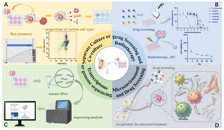
Applications of Ascites-Derived Organoids. Tumor organoids derived from clinical specimens serve as the core model supporting four functional modules. (A) Organoid separate culture and co-culture systems, with flow cytometry applied to quantify the proportions of distinct cell subpopulations. (B) Organoid-based drug screening and radiotherapy (RT) sensitivity testing, generating dose-response curves for anticancer agents and gradient radiation doses. (C) Genome/transcriptome sequencing and bioinformatic analysis using nucleic acids extracted from tumor organoids. (D) Microfluidic organoid chips recapitulating the tumor microenvironment (TME) — including cancer cells, cancer-associated fibroblasts (CAFs), macrophages and extracellular matrix (ECM) — for physiologically relevant drug screening. Abbreviations: RT, radiotherapy; TME, tumor microenvironment; CAF, cancer-associated fibroblast; ECM, extracellular matrix; Gy, Gray.
1.1.2 Influencing Factors
It is worth critical note that the reported establishment success rates of MADOs cover an extremely wide range of 33% to 100%. However, most primary studies fail to systematically dissect the drivers behind these substantial discrepancies, which has severely impaired experimental reproducibility across the field. The confounding clinical and technical variables accounting for such disparities can be categorized into four groups:
(1) Heterogeneity bias of clinical samples: Advanced cancer patients display dramatic interindividual differences in prior chemotherapy/intraperitoneal perfusion history, ascitic tumor cellularity, tumor differentiation grade and peritoneal metastatic burden. High-grade malignant samples exhibited higher organoid formation efficiency, potentially due to their greater proliferative capacity and adaptability [11]. Prior chemotherapy severely damages tumor cell viability and directly reduces organoid establishment efficiency.
(2) Technical variability in laboratory operations: Different laboratories adopt inconsistent protocols, including centrifugal force set at 300-500 × g, different matrices (Matrigel vs. Basement Membrane Extract (BME)), optional supplementation with autologous ascitic supernatant, and distinct processing workflows for fresh versus cryopreserved cells. Minor operational deviations are sufficient to alter culture outcomes. Treatment with erythrocyte lysate enhances tumor cell purity, while adding ascites supernatant provides natural growth factors. Optimizing centrifugation parameters (300-500 × g) reduces cellular damage [2, 3, 6, 13, 18, 19].
(3) Inconsistent criteria for successful culture: Some studies simply define success as microsphere formation, while others require stable passaging for at least five generations and valid readouts in drug sensitivity assays. Loose assessment criteria artificially inflate reported success rates.
(4) Limited sample sizes of clinical cohorts: Almost all single-center cohorts enroll fewer than 50 patients; for example, Yu et al. [11] enrolled 32 patients with gastrointestinal cancers (of whom 24 were successfully cultured and 14 were clinically evaluable). Small cohorts inherently generate statistical fluctuations, and large-scale multicenter cohorts for data calibration remain absent.
This wide range of reported establishment success rates leads to notable methodological drawbacks. Culture protocols and drug susceptibility datasets generated by different laboratories cannot be cross-compared, hindering the formulation of unified laboratory standards. In addition, researchers tend to selectively exclude samples with low establishment rates, resulting in publication bias and underestimating the real-world practical ceiling of MADOs for patients with refractory advanced malignant ascites.
1.2 Progress in Standardising Culture Techniques
In view of the biological characteristics of different tumor types (e.g., cellular heterogeneity, microenvironment dependence, proliferation rate, etc.), researchers have developed a series of optimized organoid culture protocols tailored to specific tumors, with the core focus on tumor cell adaptability and experimental reproducibility. The key technical framework includes efficient isolation and purification of tumor cells, screening of biocompatible matrices, personalized medium formulations supplemented with tumor type-specific growth factors/cytokines, and a rigorous quality control system. For example, the culture of ovarian cancer organoids relies on specific growth factors, whereas the medium for gastrointestinal cancer organoids needs to balance cell proliferation and phenotype maintenance [2, 4, 20]. The establishment of standardized protocols has not only markedly improved the culture success rate of tumor samples from different sources, but also provided essential support for the comparability of results across studies and institutions [2, 6, 14, 17, 21, 22]. Detailed culture key points are shown in Table 2.
Key Technical Points for Culturing Various Cancer Organoids
| Tumor Type | Core Culture Technology | Culture Period | Quality Control Key Points | References |
|---|---|---|---|---|
| Ovarian Cancer | BME matrix Growth factors + ROCK inhibitors DNase I | Short-term (6 days) | KRAS pathway validation Ki67/CK19/HE staining | Graham et al., 2023[2] Chen et al., 2020[4] |
| Pancreatic Cancer | Enzymatic digestion (collagenase II + Dispase II) hEGF Antioxidants | 3 days - 2 weeks (as needed) | Stable passaging (≥5 generations) KRAS/TP53 mutation verification CAFs/immune cell isolation | Tsai et al., 2018[6] |
| Gastrointestinal Cancer (Gastric, Colorectal, etc.) | Matrigel matrix RBC lysis Wnt3a supplementation | 7-14 days (passageable) | HER2/KRAS mutation verification Non-tumor cell depletion Histological consistency | Choi et al., 2023;[13] |
| Gynecologic Cancer (Endometrial Cancer, Ovarian Cancer) | Matrigel matrix TrypLE digestion Chemotherapy screening factors | Initiation: 2-3 days Drug testing: 7-10 days | Pathological consistency HER2 IHC Clinical drug response matching | Bi et al., 2021[14] |
| Metastatic Breast Cancer (pleural effusion; ascites subset) | BME matrix EGF/FGF Y-27632 | 7-21 days (passageable) | ER/PR/HER2 verification PI3K/AKT1 mutation detection Ki67 proliferation assay | Önder et al., 2023[18] |
| Colorectal Cancer (MSI-H Type) | Matrigel embedding Noggin/EGF/Y-27632 | 7-14 days (15 passages) | MSI/MMR validation Primary tumor mutational signature retention | Küçükköse et al., 2022[17] |
| Ovarian Cancer (PDO Model) | Serum-free medium Fresh non-cryopreserved cells ErSO screening | Drug testing: 3 days | AlamarBlue viability assay UPR pathway validation; Clinical sensitivity matching | Duraki et al., 2025[22] |
Abbreviations: BME, Basement Membrane Extract; ROCK, Rho-associated coiled-coil-containing kinase; DNase I, Deoxyribonuclease I; hEGF, human epidermal growth factor; RBC, red blood cell; Wnt3a, Wingless-type MMTV integration site family member 3A; TrypLE, Trypsin-like enzyme; EGF, epidermal growth factor; FGF, fibroblast growth factor; MSI-H, microsatellite instability-high; MMR, mismatch repair; UPR, unfolded protein response
Drugs and Validation Assays for Organoid-Based Drug Screening
| Tumor Type | Drug Sensitivity Testing Methods | Drugs Used | References |
|---|---|---|---|
| Gastrointestinal Cancer (Colorectal, Gastric, Gallbladder) | Cell viability assay (CellTiter-Glo/alamarBlue) IC₅₀ calculation Clinical efficacy validation | 5-FU, oxaliplatin, cisplatin, irinotecan, paclitaxel, raltitrexed, gemcitabine | Yu et al., 2025[11] |
| Gastrointestinal Cancer (Gastric) | Cell viability assay (CCK-8) Organoid area analysis Genomic consistency validation | Oxaliplatin, 5-FU, cisplatin, docetaxel, irinotecan, paclitaxel, epirubicin | Li et al., 2019[5] |
| Ovarian Cancer (High-grade Serous) | Cell viability assay (ATP luminescence/CellTiter-Glo 3D) Growth rate analysis RNA-seq pathway validation | Carboplatin, paclitaxel, MK-1775 (Wee1 inhibitor), CB-5083 (p97 inhibitor), sorafenib | Chen et al., 2020[4] |
| Cell viability assay (CellTiter-Glo/alamarBlue) Synergy analysis (SynergyFinder) Western blot pathway validation | Carboplatin, olaparib, niraparib, cobimetinib (MEK inhibitor), trametinib (MEK inhibitor) | Tomas et al., 2026[34] Nguyen et al., 2023[37] | |
| Ovarian Cancer | Cell viability assay (CellTiter-Glo 3D) Lipid peroxidation detection Ferroptosis marker analysis | CAY10566 (SCD1 inhibitor), sc26196 (FADS2 inhibitor), cisplatin, ferrostatin-1 | Xuan et al., 2022[20] |
| Cell viability assay (CellTiter-Glo 3D) Live-cell imaging (Calcein AM) In vivo peritoneal metastasis model validation | MF438 (SCD1 inhibitor), oleic acid | Guo et al., 2025[38] | |
| Cell viability assay (ATP luminescence) Single-drug sensitivity assessment | Carboplatin | Fashemi et al., 2023[33] | |
| Colorectal Cancer | Cell viability assay Dose-response curve analysis Rare mutation targeted drug validation | Afatinib (EGFR-TKI), osimertinib (EGFR-TKI) | Kondo et al., 2025[23] |
| Gynecologic Cancer (Ovarian, Endometrial) | Cell viability assay Chemotherapy resistance prediction Second-line regimen screening | Carboplatin, cisplatin, paclitaxel, bevacizumab, gemcitabine, topotecan | Bi et al., 2021[14] |
Abbreviations: IC₅₀, half-maximal inhibitory concentration; ATP, adenosine triphosphate; CCK-8, Cell Counting Kit-8; EGFR-TKI, epidermal growth factor receptor-tyrosine kinase inhibitor
The culture of various tumor organoids has been standardized and optimized in accordance with their unique biological characteristics, with distinct core highlights: Graham et al. [2] optimized the cell isolation protocol for multi-source ovarian cancer samples to ensure phenotypic fidelity in short-term culture; Duraki et al. [22] established an ovarian cancer PDO model, which enables rapid 3-day drug sensitivity testing using fresh ascites samples and caters to the demands of novel therapeutic screening. This platform eliminates the requirement for serial passaging, thereby circumventing genomic drift and clonal selection to the maximum extent arising from prolonged serial expansion. The pancreatic cancer protocol developed by Tsai et al. [6] balances sample compatibility and microenvironment simulation, allowing simultaneous isolation of relevant stromal and immune cells to support the construction of complex models.
In the field of gastrointestinal cancer, Choi et al. [13] focused on solving sample purification issues, while Küçükköse et al. [17] optimized the protocol for MSI-H colorectal cancer to guarantee model reliability for immunotherapy-related research. The gynecologic cancer protocol proposed by Bi et al. [14] emphasizes clinical adaptability, with drug sensitivity results that can directly provide a basis for personalized treatment; the metastatic breast cancer protocol established by Önder et al. [18] ensures the model aligns with the characteristics of clinical metastatic lesions through rigorous validation of receptor status and gene mutations.
Overall, all these protocols follow the triple quality control principle of morphology-molecular profile-function, ensuring the biological consistency between organoids and primary tumors as well as experimental reproducibility [14]. These standardized protocols not only highlight tumor-type specificity but also meet the demands of clinical translation, laying a solid foundation for the application of organoid technology in precision medicine and basic research [22].
2. Biological characteristics and tumor modelling capacity of ascites organoids
2.1 Consistency with Primary Tumors
Ascites organoids have been successfully established from multiple malignancies including gastric, ovarian, and colorectal cancers, exhibiting histological and molecular characteristics highly consistent with primary tumors [3, 11, 23]. At the histological level, immunohistochemical features of breast cancer ascites organoids align with primary tumor tissues [13]. At the molecular level, gastric cancer ascites organoids can mimic the microenvironment of peritoneal metastases in co-culture systems, preserving tumor heterogeneity and drug response characteristics [3]. Ovarian cancer ascites organoids can form within a short period (within 6 days) and maintain the tumor's transcriptomic characteristics, including key biological processes such as proliferation, epithelial-mesenchymal transition, and KRAS signalling pathways [4]. Single-cell transcriptomic analysis revealed that ascites organoids retain tumor heterogeneity, encompassing proliferative and non-proliferative cell subpopulations, with this heterogeneity undergoing dynamic changes under chemotherapy stress [8]. Furthermore, ovarian cancer ascites organoids exhibit high similarity to primary tumors in metabolic characteristics (e.g., upregulation of oxidative phosphorylation) and gene expression profiles (e.g., activation of the MYC signalling pathway), providing a model for investigating chemoresistance mechanisms [8]. However, a key unresolved controversy in the field is whether long-term passaging of MADOs preserves complete intratumoral heterogeneity: some studies report stable clonal diversity after multiple passages, while others observe progressive depletion of rare subclones and genomic drift [24, 25], highlighting the need for case-by-case validation based on culture duration and research objectives.
2.2 Capacity to Simulate the Tumor Microenvironment
Ascites-derived organoids not only retain the inherent biological characteristics of tumor cells but also recapitulate the interactions between tumors and the microenvironment via co-culture systems. When gastric cancer ascites-derived organoids are co-cultured with peritoneal mesothelial cells, soluble factors secreted by mesothelial cells markedly suppress organoid proliferation and induce paclitaxel resistance in the organoids, directly illustrating the molecular mechanism underlying tumor microenvironment-mediated chemoresistance [3]. In ovarian cancer models, Akt-activated endothelial cells form "vascular spheres" that promote tumor peritoneal implantation via Notch3/Jagged1 signalling, a phenomenon validated in patient ascites [26]. For pancreatic cancer, researchers integrated tumor-associated fibroblasts (CAFs) and lymphocytes via a co-culture system, establishing the first 3D microenvironment model incorporating tumor-stromal-immune tripartite components. They observed myofibrillar activation of CAFs and tumor-dependent T-cell infiltration, highly mimicking the complex composition of the in vivo tumor microenvironment [6]. Furthermore, co-culture systems integrating ascites organoids with immune cells (such as T cells and macrophages) have been established to evaluate immunotherapy efficacy, providing a reliable platform for elucidating tumor-immune cell interactions [6, 27].
2.2.1 Emerging Mechanisms: EV-Mediated Crosstalk and Mechanobiology
Recent advances highlight two understudied aspects of MADO microenvironment simulation: extracellular vesicle (EV) communication and mechanobiology. Ascites is enriched with EVs secreted by tumor cells, stromal cells, and immune cells, which serve as key mediators of intercellular communication. Native ascites EVs are enriched with tumor-derived miRNAs including miR-1246 and miR-1290, which promote ovarian cancer cell invasion by targeting RORα [28]. MADO models provide a tractable ex vivo platform in which the functional effects of these ascites-derived EV cargos can be dissected; however, direct evidence that EVs released by cultured MADOs in vitro faithfully recapitulate the cargo of native ascitic EVs remains to be established. Co-culture experiments demonstrate that tumor-derived EVs in ascites modulate stromal cell activation and immune suppression, contributing to chemoresistance and metastasis [29].
Additionally, the biophysical characteristics of the ascites microenvironment (e.g., fluid shear force, osmotic pressure, extracellular matrix stiffness) play a critical role in tumor progression but are rarely recapitulated in conventional MADO cultures. The peritoneal cavity exhibits dynamic fluid flow (0.1-1 mm/s) and viscous stress, which regulate tumor cell adhesion, EMT, and drug uptake [30, 31]. Recent organoid-on-chip models have begun to integrate these mechanical cues: microfluidic perfusion systems simulating ascites fluid flow enhance the biomimicry of MADO models, revealing that shear stress promotes tumor cell dissemination by upregulating integrin β1 expression [32]. These findings highlight the need to incorporate biophysical parameters into future MADO culture systems to better replicate the in vivo microenvironment.
3. Core Application Value of Ascites Organoids
3.1 Drug Screening and Personalized Treatment Guidance
3.1.1 Drug Screening
Multicellular spheroids or tumor cells isolated from malignant ascites can be induced and cultured into organoids within a short period and used for drug sensitivity testing, providing rapid and reliable references for clinical treatment decision-making [5, 11, 14]. For instance, drug sensitivity testing of ovarian cancer ascites-derived organoids can be completed within 7-10 days, significantly shortening the cycle of conventional drug sensitivity assays [4, 33]; the culture success rate of ascites-derived organoids from gynecologic cancers (ovarian cancer, endometrial cancer) is as high as 85.7% (6 out of 7 ascites samples), and drug sensitivity testing can be completed within 7-10 days after sample collection, supporting the timely formulation of individualized regimens [14]. However, given the small sample size, these findings should be interpreted as preliminary and warrant validation in larger cohorts. Ovarian cancer ascites organoids have not only successfully predicted patients' acquired resistance to trastuzumab but also screened out alternative second-line treatments such as PARP inhibitors [14, 34].
In predicting clinical drug responses, ascites-derived organoids demonstrate excellent accuracy: the prediction accuracy of clinical chemotherapeutic efficacy for gastrointestinal cancer ascites organoids reaches 78.57%-85.71% (Kappa value 0.571-0.714) [11]. Nevertheless, these predictive values must be interpreted with caution, as they are derived from small, single-center retrospective cohorts with inherent methodological limitations that may overestimate real-world performance.
First, small sample sizes and culture-dependent selection bias limit generalizability. The cohort reporting these accuracy values included only 32 malignant ascites samples, of which 24 were successfully cultured and 14 had evaluable matched clinical outcomes [11]. Most MADO validation studies enroll 20-35 patients, and systematically exclude samples with paucicellular ascites or indolent tumor clones that fail to proliferate in vitro. This growth-dependent selection skews predictive performance toward metabolically active tumors, rather than reflecting the full spectrum of clinical peritoneal metastasis [35, 36].
Second, retrospective designs introduce verification bias. Nearly all current MADO predictive studies match organoid sensitivity to clinical outcomes retrospectively, with response thresholds often optimized post hoc rather than predefined in a blinded prospective cohort [35, 36]. To date, no prospective, blinded trial has demonstrated that MADO-guided therapy improves progression-free or overall survival compared with standard of care.
Third, pre-analytical sample heterogeneity undermines representativeness. Ascites fluid contains variable proportions of tumor cells, immune cells, mesothelial cells, and spatially distinct tumor clones from multiple peritoneal implants. During organoid establishment, long-term 3D culture preferentially selects for rapidly proliferating epithelial clones, while slow-cycling drug-resistant subclones and stromal components are gradually lost [35, 36]. This pre-analytical selection means the final organoid population may not faithfully represent the full clonal architecture of the original tumor.
Nevertheless, predictive assays using organoids yield both false-negative and false-positive results. One study documented that the specificity of 5-FU response prediction reached 85.71%, yet the corresponding sensitivity was comparatively low, indicative of prevalent false negatives [11]. This high false-negative rate carries clinical consequences: a negative organoid result may lead clinicians to incorrectly withhold a potentially effective therapy. This limitation stems from the failure of organoid models to faithfully recapitulate intricate in vivo drug metabolic pathways and immune surveillance networks [11, 35]. Unlike the in vivo setting, standard MADO cultures lack functional drug-metabolizing enzyme systems, stromal metabolic niches, and endogenous immune cells, which can alter drug bioavailability and produce artificially resistant readouts, particularly for immunomodulatory and stroma-targeted agents [35, 36].
More importantly, the core gap in current evidence is not merely methodological, but translational: few studies have directly demonstrated that predictions derived from MADOs can genuinely alter patients' clinical outcomes or improve survival. Therefore, the level of evidence supporting the clinical predictive value of MADOs remains insufficient, and large-scale prospective, blinded clinical trials are needed for further validation.
3.1.2 Personalized Treatment Guidance
Ascites-derived organoids exhibit significant heterogeneous responses to standard chemotherapeutic agents, a property that provides core support for the formulation of individualized treatment regimens [5, 11, 14, 37]. For example, in high-grade serous ovarian carcinoma, the combination of MEK inhibitors (cobimetinib and trametinib) with carboplatin produces a synergistic cytotoxic effect on NF1-mutant organoids. By inhibiting the RAS-ERK pathway, this combination regimen downregulates genes such as c-MYC and FOXM1, thereby providing a novel individualized therapeutic strategy for patients with platinum-resistant ovarian cancer [37]; SCD1/FADS2 inhibitors (CAY10566, SC26196) induce ferroptosis and disrupt redox homeostasis, synergistically inhibiting peritoneal metastasis of ovarian cancer when combined with cisplatin, while reversing platinum resistance. This strategy is suitable for ovarian cancer patients with aberrant lipid metabolism [20]; the SCD1 inhibitor MF438 can also block epithelial-mesenchymal transition (EMT) by suppressing the TGFβ-Smad3 signaling pathway, significantly inhibiting intraperitoneal dissemination of ovarian cancer. It can be used for individualized intervention in patients with high metastatic risk [38]. Ascites-derived organoids also facilitate the discovery of novel therapeutic targets. For example, the Wnt signaling pathway exerts a key regulatory role in peritoneal metastasis of gastric cancer, providing new strategies for targeted intervention in patients with advanced gastric cancer with aberrant Wnt pathway activation [19]. For patients with rare mutations, ascites organoids show unique value: ascites-derived organoids from colorectal cancer patients with EGFR kinase domain duplication (EGFR-KDD) have confirmed their sensitivity to EGFR tyrosine kinase inhibitors (such as afatinib and osimertinib), providing personalized therapeutic options for such patients lacking standard treatment regimens [23]. Non-proliferative cell subpopulations remaining after chemotherapy highly express genes associated with oxidative phosphorylation (OXPHOS), suggesting that metabolic phenotypic switching is critical for the survival of drug-resistant cells [1, 8].
These findings indicate that, as an efficient in vitro model, ascites-derived organoids can accurately recapitulate the histological features, genomic characteristics and drug response heterogeneity of tumors, successfully covering various metastatic cancer types including ovarian cancer, gastrointestinal cancer and gynecologic cancer, thus serving as a core decision-making tool for individualized therapy. They hold important clinical translational value especially for advanced cancer patients who have failed standard treatments.
3.2 Research into Resistance Mechanisms
Ascites organoids provide a powerful tool for deciphering the molecular mechanisms of chemotherapy resistance, revealing multiple pathways of resistance. A key unresolved controversy in the field is the relative contribution of intrinsic tumor cell alterations versus microenvironment-mediated factors to chemoresistance: some studies emphasize that metabolic reprogramming (e.g., OXPHOS upregulation) is the primary driver [39], while others highlight the dominant role of microenvironmental crosstalk (e.g., mesothelial cell-tumor cell interactions) [3, 40]. This discrepancy may be attributed to tumor-type specificity—for example, ovarian cancer resistance is more strongly associated with microenvironment factors, whereas gastric cancer resistance is driven by intrinsic metabolic changes [41].
3.2.1 Immune Evasion and Microenvironment Remodeling
Microenvironment-mediated drug resistance and metastasis are critical mechanisms driving ovarian cancer progression. In addition to the direct crosstalk between peritoneal mesothelial cells and tumor cells that mediates platinum resistance, ascites-derived small extracellular vesicles (sEVs) can significantly promote the migration, invasion, and peritoneal metastasis of ovarian cancer cells through multiple signaling mechanisms, including TGFβ-related pathways and other microenvironmental reprogramming mechanisms. Additionally, mesothelial cell-derived factors such as FN1/AKT signaling further contribute to platinum resistance and metastatic progression [42-46]. Intrinsic molecular alterations in tumor cells also govern resistance regulation. For instance, tumor-derived UBR5 has been shown (in ovarian cancer cell lines and murine models) to enhance tumor stemness and resistance by modulating β-catenin signaling and recruiting immunosuppressive macrophages [47]; whether this mechanism operates similarly in primary MADO models remains to be confirmed.
3.2.2 Metabolic Reprogramming
Metabolic reprogramming is also a key factor in resistance. Research indicates that in ascites-derived ovarian cancer cells, fatty acid desaturases SCD1 and FADS2 are abnormally overexpressed, promoting unsaturated fatty acid synthesis, tumor stem cell formation, and platinum resistance. Inhibiting SCD1/FADS2 disrupts cellular redox balance, induces ferroptosis, and produces synergistic antitumor effects with cisplatin [20]. Moreover, metabolite profiling has revealed that ascites-derived ovarian cancer cells contain substantially higher intracellular taurine levels than cultured counterparts. Functional studies in established two-dimensional ovarian cancer cell lines and patient-derived organoids have shown that elevated taurine activates p53, ERK, mTOR, and DNA damage response pathways, inhibiting cell proliferation and glycolysis while inducing cisplatin-protective effects that can be counteracted by mTOR/DNA damage response pathway inhibitors [48]. Whether these effects operate similarly in primary ascites-derived models remains to be confirmed.
There exists a complex crosstalk network among distinct drug resistance mechanisms. For instance, metabolic reprogramming (such as SCD1/FADS2-mediated fatty acid synthesis) not only directly induces chemoresistance, but also promotes EMT by modulating the TGFβ-Smad3 signaling pathway, thereby enhancing tumor invasiveness and drug resistance. Such multi-pathway synergistic effects represent a major cause of clinical chemoresistance.
Analyzing Mechanisms of Tumor Biology
3.3.1 Tumor Heterogeneity and Evolution
Single-cell RNA sequencing (scRNA-seq) technology has profoundly deepened our understanding of the peritoneal cavity ecosystem. Studies reveal high heterogeneity among cancer and immune cells in the ascites of gastric cancer patients with peritoneal metastasis [10]. Researchers traced highly plastic gastric cancer cell clusters capable of transitioning to a highly proliferative phenotype via an autophagy-dependent "paligenosis" programme, associated with poor prognosis [10]. In ovarian cancer, whole-genome sequencing combined with evolutionary analysis classified high-grade serous ovarian carcinomas into three evolutionary states with distinct genomic signatures, pathway activities, and therapeutic responses, outlining evolutionary trajectories that provide a novel framework for patient stratification [16].
3.3.2 Intercellular Communication Networks
By integrating single-cell RNA-seq and bulk RNA-seq data, researchers systematically mapped the interaction network between cancer-associated fibroblasts and cancer cells. They identified key ligand-receptor pairs such as THBS2/THBS3-CD47 and MDK-NCL/SDC. These interactions were enriched in the PI3K-AKT, focal adhesion, and EMT pathways and were significantly correlated with patient prognosis [9]. Co-culture experiments between ascites organoids and microenvironmental cells further confirmed that these intercellular communication pathways play a central role in tumor proliferation, migration, and drug resistance [26].
3.3.3 Characteristics of the Immune Microenvironment
Ascites organoid models reveal multi-layered mechanisms of immunotherapy resistance in peritoneal metastases. MADOs exhibit great potential for immuno-oncology research. The establishment of T cell-organoid co-culture systems enables efficient evaluation of the therapeutic efficacy of immune checkpoint inhibitors, such as anti-PD-1 and anti-PD-L1 antibodies. Furthermore, MADO models are applicable to functional testing of CAR-T cell therapy, screening and validation of neoantigens, as well as the isolation and characterization of tumor-reactive lymphocytes. Collectively, these applications constitute a critical ex vivo platform for the development of novel immunotherapeutic strategies.
In humanised mouse models of microsatellite-unstable colorectal cancer (MSI-H CRC), peritoneal metastases exhibit intrinsic resistance to PD-1/CTLA-4 inhibitors. This resistance correlates with the absence of B cells and tertiary lymphoid structures (TLS), T-cell dysfunction, and elevated levels of immunosuppressive cytokines such as IL-10 and TGF-β in ascites [17]. B-cell depletion experiments confirmed B cells are critical for the antitumor efficacy of CTLA-4 inhibitors, whilst the ascites microenvironment induces T-cell functional exhaustion via high concentrations of immunosuppressive factors [17]. Single-cell sequencing classified diffuse gastric carcinoma (DGAC) ascites into two subtypes: CDH1-deficient (DGAC1) and immune-silent (DGAC2). DGAC1 exhibits distinct molecular characteristics and a T-cell exhaustion phenotype. Animal studies confirm that Cdh1 knockout accelerates tumorigenesis and promotes immune evasion. Mechanistic investigations reveal EZH2 as a key mediator of CDH1-deficient tumorigenesis [7]. These findings provide novel targets for developing targeted immunotherapy strategies [7, 49, 50].
3.4 Biomarker Discovery and Liquid Biopsy
Ascites serves not only as a source of tumor cells but also as a liquid biopsy sample rich in tumor-derived biomolecules. MADO-based organoid technology provides a unique platform for functional validation of liquid biopsy biomarkers. Ascites-derived cell-free DNA (cfDNA) and extracellular vesicles (EVs) act as key biomarkers for tumor diagnosis and treatment response monitoring.
For patients with colorectal cancer peritoneal metastases, tumor-derived cell-free DNA (cfDNA) levels in plasma are extremely low, whereas cfDNA in ascites (or peritoneal lavage fluid) exhibits significantly higher mutated allele frequencies and copy numbers, suggesting ascites cfDNA is a superior biomarker for monitoring peritoneal spread [51]. Extracellular vesicles (EVs) in ascites also serve as important information carriers. More importantly, extracellular vesicles (EVs) secreted by in vitro-cultured MADOs have been proven to faithfully recapitulate the molecular signatures of native ascitic EVs. For example, miR-1246 and miR-1290 carried by ascitic EVs promote ovarian cancer cell invasion and migration by targeting RORα, and this biological function is recapitulated in MADO models [28].
Accordingly, MADO models serve as a robust platform to functionally validate ascites-derived liquid biopsy biomarkers and further dissect their mechanistic roles in tumor progression.
3.5 Development of Novel Therapeutic Strategies
The ascites organoid platform accelerates the evaluation and optimization of novel therapies across multiple domains, including targeted therapies, immunotherapies, nanomedicines, and metabolic interventions.
In targeted therapies, inhibitors targeting the WNT signalling pathway (gastric cancer peritoneal metastasis), SCD1/FADS2 (ovarian cancer), and EZH2 (diffuse gastric cancer) have demonstrated efficacy validated through organoid models [7, 19, 20]; in immunotherapy, co-culture systems of colorectal cancer organoids with autologous immune cells have been used to show that combining PD-1 blockade with standard chemotherapy enhances antitumor efficacy beyond either modality alone [27]. Nanoparticles loaded with EP2/EP4 antagonists can reverse prostaglandin E2-induced functional suppression of dendritic cells, restoring antitumor immunity [52]. Nanodrug development also benefits from ascites organoid models, such as imatinib encapsulated within bola-amphiphilic dendritic polymers (Bola/IM) effectively inhibiting tumor stemness and metastasis in ovarian cancer organoids by targeting the β-catenin/HRP2 axis [53].
Additionally, CD206-targeted mannose-modified nanoparticle platforms have been shown to selectively deliver siRNA to M2-type tumor-associated macrophages in ovarian cancer models, reprogramming the immunosuppressive tumor microenvironment and reducing ascites volume [54]. Furthermore, the atypical UPR (aUPR) hyperactivator ErSO induced necrotic death in all seven ovarian cancer ascites organoids, activating macrophages through damage-associated molecular patterns (DAMPs) release to exert both direct cytotoxic and immunomodulatory effects [22]; inhibiting the palmitoyltransferase ZDHHC12 significantly enhances ovarian cancer organoid sensitivity to cisplatin by increasing reactive oxygen species (ROS) levels [1].
For rare tumors such as fibrolamellar hepatocellular carcinoma (FLC), solid tumor derived organoid models have shown promising preclinical value, with DNAJB1::PRKACA identified as a key therapeutic target [55]. However, malignant ascites derived organoid (MADO) models for FLC have not yet been reported, and drug screening data based on ascites derived models remain scarce for this rare malignancy.
4. Technical Challenges and Future Directions
4.1 Challenges
4.1.1 Defects in Microenvironment Biomimetic Simulation
Although ascites-derived organoids possess promising application prospects, they present notable deficiencies in microenvironment biomimicry, which severely restricts the authenticity and applicability of the models. Current models are incapable of fully recapitulating the tripartite interactions among tumor cells, fibroblasts and immune cells, making it difficult to accurately replicate the complex cellular communication network in vivo [14]. The conventional MADO culture system cannot sustain long-term stable retention of various stromal and immune components, including T cells, NK cells, fibroblasts, and endothelial cells. In addition, it tends to induce the loss of stromal components of primary tumors in organoids, failing to truly recapitulate microenvironment-dependent tumor biological behaviors [6]. Meanwhile, current mainstream static culture systems have intrinsic limitations. They fail to recapitulate the complex structure of the in vivo extracellular matrix (ECM) and cannot simulate key mechanical cues such as fluid shear force. Overall, MADO models present insufficient biomimicry. When utilized for mechanistic research that relies heavily on intact microenvironments, these models tend to overestimate the consistency between the in vitro model state and the authentic in vivo physiological state.
4.1.2 Practical Limitations of Culture Systems
The technical system for clinical and scientific research applications of ascites-derived organoids still has multiple practical limitations. Firstly, the standard MADO drug-sensitivity assay requires approximately 3-7 days from sample receipt to result output, which is substantially faster than PDX models (months) or solid-tumor PDOs (2-3 weeks). Nevertheless, in patients with rapidly progressive peritoneal carcinomatosis and deteriorating performance status, even a 7-day turnaround may compress the window for timely regimen adjustment [6, 18]. This clinical urgency motivates the development of no-passage rapid platforms (e.g., the 3-day assay of Duraki et al. [22]) and AI-based predictive models that further shorten decision time. Secondly, clinically derived samples exhibit high heterogeneity, resulting in substantial variations in the drug response of organoids. For instance, the IC₅₀ values of gastric cancer ascites-derived organoids against irinotecan vary by up to two orders of magnitude across patient-derived lines, markedly increasing the difficulty of result interpretation [5]. Thirdly, there is a lack of a unified standardized culture system in the industry. Medium formulations, cell passaging protocols, and quality control criteria vary substantially across different laboratories. This not only reduces the comparability of research findings among different studies, but also hinders the progression of multicenter MADO clinical trials. Consequently, this technology is difficult to integrate into hospital routine clinical systems and cannot be standardized and widely applied [17, 22].
4.1.3 Risks of Genomic Instability during In Vitro Passaging
Beyond the aforementioned limitations, serial passaging of MADOs in vitro poses two critical risks, namely genomic drift and clonal selection, which impair the organoids' capacity to faithfully recapitulate the intratumoral heterogeneity and genomic landscapes of primary tumors; short-term and long-term culture encounter distinct challenges in maintaining tumor clonal features. Short-term culture (3-10 days) tailored for immediate clinical drug screening demands minimal passaging and maintains robust genomic stability, accurately recapitulating the clonal architecture of patient-derived primary lesions [25]. By contrast, prolonged expansion conducted for mechanistic research imposes ex vivo selective pressure, under which fast-proliferating subclones gradually outcompete other cell populations. This process drives progressive genomic drift and results in the depletion of rare tumor subclones and loss of global intratumoral heterogeneity, ultimately yielding genomic discrepancies between cultured MADOs and in vivo patient neoplasms [24]. Therefore, experimental designs should stratify culture lengths based on research purposes: short-term incubation is ideal for retaining native tumor heterogeneity, whereas long-term serial passaging studies must account for clonal bias stemming from clonal skewing. When conducting long-term studies, it is essential to regularly assess the genomic integrity of organoids and compare them with the original tumor samples.
4.1.4 Bottlenecks in Clinical Translation and Application
The translation of ascites-derived organoid models from basic research to clinical application still faces prominent barriers, including clinical, economic, and regulatory challenges. Clinically, current validation studies are limited by small sample sizes and insufficient cohort representativeness (e.g., Yu et al.[11] enrolled 32 patients (24 successfully cultured, 14 clinically evaluable)), and the long-term consistency between in vitro drug sensitivity results and actual clinical efficacy remains insufficiently validated by prospective trials. Economically, high culture costs (≈$500-$1000 per sample) and the need for specialized laboratory infrastructure restrict widespread clinical adoption, particularly in resource-limited settings. Regulatorily, MADO-based testing platforms require CLIA/CAP certification to guarantee consistent quality control, and robust clinical decision support frameworks are lacking to guide the integration of MADO results into routine clinical practice.
4.2 Future Directions
4.2.1 Optimizing Culture Systems to Bridge the Gap in Microenvironment Biomimicry
To address the incomplete microenvironment simulation, low biomimetic fidelity of static systems, and prolonged culture cycles in current MADO models, future efforts must prioritize the optimization of culture systems. By supplementing matrix components, optimizing conditioned media, or introducing microfluidic technologies to upgrade traditional culture modes, it is possible to effectively preserve the intact tumor microenvironment and recapitulate in vivo fluid dynamics and mechanical characteristics. This approach not only shortens the model culture cycle but also resolves the core issues of insufficient biomimicry and poor timeliness inherent in traditional systems [21, 26]. Meanwhile, organ-on-a-chip systems can precisely simulate in vivo fluid flow and mechanical forces, providing a superior in vitro model for studying tumor-vascular interactions and further enhancing the physiological relevance of the models.
4.2.2 Establishing Standardized Frameworks to Consolidate the Foundation for Technical Application
To tackle the challenges of inconsistent culture standards across laboratories, significant interference from sample heterogeneity, and poor comparability of research findings, it is essential to establish a multi-center standardized database. By systematically integrating functional assay data of ascites-derived organoids, high-throughput sequencing data, and comprehensive clinical case information, a unified data standard and quality control system can be formed. This will effectively resolve the issue of excessive discrepancies and lack of cross-comparability among different studies. Furthermore, it will provide core data support for model optimization, big data analysis, and the advancement of multi-center clinical trials, thereby facilitating the standardized integration of this technology into routine clinical practice [4, 34].
4.2.3 Developing Dynamic Monitoring Technologies to Mitigate the Risks of Genomic and Clonal Bias
To address issues such as genomic drift, clonal selection, subclone loss, and decreased intratumoral heterogeneity induced by long-term in vitro passaging, a comprehensive dynamic monitoring system must be constructed. By leveraging cutting-edge tools such as single-cell sequencing and genetically encoded fluorescent biosensors, researchers can track the evolutionary dynamics of subclones within ascites organoids under drug pressure in real time. This allows for the precise capture of genomic variations and clonal selection processes during in vitro culture, effectively circumventing the distortion of experimental results caused by prolonged passaging and ensuring the accurate recapitulation of the primary tumor's genomic features and heterogeneity [8, 56, 57]. Additionally, the application of multifunctional biosensors (e.g., HyPer) enables the real-time dynamic monitoring of organoid metabolic states and signaling pathway activities, further improving the precision of mechanistic studies [57].
4.2.4 Integrating Emerging Technologies to Advance Clinical Translation and Precision Therapy
Relying on multi-technology integration and innovation, we can break through the barriers to the clinical translation of ascites organoids and expand the boundaries of model applications. At the technological innovation level, organoids derived from circulating tumor cells (CTCs) enable continuous dynamic monitoring of tumor progression [56]; artificial intelligence (AI) predictive models can rapidly screen drug sensitivity based on gene expression profiles, significantly shortening the time required for clinical decision-making [6, 18]; spatial transcriptomics can elucidate the internal gene expression profiles and intercellular communication mechanisms within MADOs at single-cell resolution, deepening the mechanistic study of the tumor microenvironment; the combination of digital pathology and AI can achieve automated, high-precision analysis of organoid morphology and drug responses. At the clinical application level, personalized clinical trials based on ascites organoids will become a mainstream direction. By utilizing organoid drug sensitivity results to guide patient enrollment and treatment selection, the success rate of clinical trials can be effectively improved [34, 58]. Furthermore, constructing immune cell-organoid co-culture models will allow for in-depth investigation into the mechanisms of immunotherapy resistance and the screening of novel combination strategies—such as chemotherapy combined with targeted metabolic agents, or immune checkpoint inhibitors combined with cytokines—providing new therapeutic options for patients with peritoneal metastatic cancer [6, 17, 59].
5. Discussion
As an emerging tumor model, ascites organoids have become a pivotal bridge linking fundamental research and clinical application due to their core advantage of highly preserving primary tumor characteristics. They demonstrate immense potential in elucidating tumor biological mechanisms, predicting treatment responses, and developing novel therapies. Research has progressed from early model establishment to leveraging multi-omics technologies and exploring tumor complexity, providing crucial support for precision treatment of advanced cancers, particularly peritoneal metastatic cancers [3, 20, 56].
Current research confirms that ascites organoids serve as potent in vitro tools for reproducing tumor heterogeneity, evolutionary dynamics, and drug responses, with their drug sensitivity predictions exhibiting high concordance with clinical outcomes [11, 14]. Through single-cell and spatial omics technologies, researchers have not only unravelled intricate interaction networks between cancer cells, stromal cells, and immune cells within the ascites microenvironment, but also uncovered novel resistance mechanisms involving mesothelial cells - tumor interactions, immunosuppressive microenvironments, and metabolic reprogramming. This has identified novel resistance mechanisms and potential therapeutic targets such as the PGE2-EP2/4 axis, EZH2, and OXPHOS [3, 7, 8, 52]. Concurrently, ascites liquid biopsy (cfDNA, EVs) demonstrates superior potential to plasma in monitoring and diagnosing peritoneal metastasis [28, 51]. Furthermore, specific intervention strategies targeting immunosuppression (e.g., metabolic targeting, immunomodulators) [52] provide direct experimental evidence for combination therapies. Additionally, ascites organoid models have deepened our understanding of peritoneal metastasis mechanisms (metabolic adaptation, immune evasion) [8, 17, 20], laying the groundwork for developing combination strategies integrating chemotherapy with targeted or immunotherapy [8, 14].
Supported by abundant in vitro pharmacodynamic data, a series of prospective cohort studies and clinical trials have been launched globally to explore the translational potential of MADOs in guiding individualized anti-tumor therapy. Current research is dominated by small-scale, exploratory single-center cohorts. Cohorts enrolling patients with malignant ascites secondary to gastrointestinal and gynecologic cancers have confirmed that organoid drug sensitivity profiles can rapidly guide clinical regimen formulation, and the OVAREX [58] trial is an ongoing prospective observational study designed to establish ex vivo ovarian cancer models and evaluate their feasibility and predictive value for therapeutic response stratification; completed results are not yet available. Multiple Phase II randomized controlled trials (RCTs) are currently ongoing. However, large multicenter Phase III confirmatory trials remain scarce, and the lack of standardized culture protocols hinders broad clinical adoption.
Unlike the traditional empirical treatment strategy that only depends on radiological imaging and static genomic sequencing, MADOs with minimally invasive sampling and rapid drug sensitivity assays can be integrated into the full clinical decision-making workflow for patients with advanced malignancies complicated by malignant ascites. At initial diagnosis, the 3-day rapid PDO platform allows clinicians to screen optimal first-line combination regimens. For patients with recurrent chemoresistance or tumors carrying rare mutations, serially cultured MADOs aid the identification of second-line targeted agents. Furthermore, MADOs facilitate the development of combination therapies and subject recruitment for novel clinical trials. By transforming therapeutic decision-making from static genomic prediction to functional pharmacodynamic assessment, MADOs fundamentally streamline the decision-making process of multidisciplinary tumor boards.
Nevertheless, the standardization and clinical translation of malignant ascites-derived organoids (MADOs) are still confronted with multiple technical hurdles, including sample heterogeneity, limited culture success rates, and incomplete recapitulation of the tumor microenvironment [13, 14]. From a technical perspective, lengthy assay turnaround time, variability in sample quality, and elevated culture failure rates constitute primary obstacles. Both cost and regulatory considerations remain key barriers to clinical adoption, as discussed in Section 4.1.4. Accordingly, future development should focus on core directions: optimizing culture systems to preserve more immune components [2, 6]; developing artificial intelligence predictive models to shorten culture cycles and dynamic co-culture systems integrating multi-cell components [4, 6]; integrating multi-omics analysis, functional validation and clinical information, refining personalized drug sensitivity screening platforms [18, 19]; and advancing organoid-guided clinical trial design, incorporating innovative therapies such as nanocarriers and immunotherapy [17, 52, 53, 59], accelerating their adoption as standard tools for precision medicine.
With ongoing technological advances, peritoneal ascites organoids hold promise for deepening our understanding of peritoneal metastasis mechanisms and driving novel targeted drug development [19, 38]. This may ultimately enable personalized treatment guidance for advanced cancer patients, improving survival outcomes and quality of life [11, 14].
6. Conclusion
As an emerging tumor model, ascites-derived organoids exhibit irreplaceable value in preserving primary tumor characteristics, recapitulating microenvironmental crosstalk, and guiding personalized therapy. The improved successful establishment rate and standardization of culture techniques have laid a foundation for bridging basic research and clinical application. Using ascites-derived organoid models, researchers have uncovered key molecular pathways underlying tumor heterogeneity, drug resistance, and immune escape, identified a panel of potential biomarkers and therapeutic targets, and accelerated the development of novel therapies.
However, several critical issues must be resolved to achieve widespread clinical application of ascites-derived organoids, including sample heterogeneity, standardized culture, complete microenvironmental recapitulation, and clinical validation. In the future, with the integration of multi-omics profiling, artificial intelligence (AI) and microfluidic technologies, alongside optimized culture frameworks and standardized operating protocols as well as large-scale prospective clinical trials, MADOs will not only become routine tools for precision therapeutic management of advanced cancers, but also thoroughly transform individualized treatment strategies for patients with peritoneal metastasis based on the ever-expanding body of clinical research, thereby effectively boosting patients' survival prognosis and quality of life.
Acknowledgements
Funding information
This research was supported by Natural Science Foundation of Fujian Province (2026J0012074, 2023J011269), Fujian Provincial Health Science and Technology Plan project (2023QNA087), Fujian Medical University Sailing Fund Project (2022QH 1166).
Author contributions statement
YD conceived the idea. TL wrote the review with supervision from YW, FK, QX and YD provided critical feedback. TL made the figures. All authors contributed to the final manuscript. YD is the guarantor of the review.
Artificial Intelligence (AI)
During the preparation of this work, AI was used solely for basic grammar and spelling correction. No AI was used for content generation, data analysis, interpretation, or manuscript structuring. The authors take full responsibility for the accuracy, integrity, and originality of the content.
Competing Interests
The authors have declared that no competing interest exists.
References
1. Zhang X, Liao X, Wang M. et al. Inhibition of palmitoyltransferase zdhhc12 sensitizes ovarian cancer cells to cisplatin through ros-mediated mecha-nisms. Cancer Sci. 2024;115:1170-83
2. Graham O, Rodriguez J, van Biljon L. et al. Generation and culturing of high-grade serous ovarian cancer patient-derived organoids. J Vis Exp. 2023
3. Uematsu H, Shimizu S, Onuma K. et al. Interaction between gastric cancer organoids and peritoneal mesothelial cells suppresses the growth of gastric cancer organoids and induces drug resistance. Hum Cell. 2025;39:8
4. Chen H, Gotimer K, De Souza C. et al. Short-term organoid culture for drug sensitivity testing of high-grade serous carcinoma. Gynecol Oncol. 2020;157:783-92
5. Li J, Xu H, Zhang L. et al. Malignant ascites-derived organoid (mado) cul-tures for gastric cancer in vitro modelling and drug screening. J Cancer Res Clin Oncol. 2019;145:2637-47
6. Tsai S, McOlash L, Palen K. et al. Development of primary human pancre-atic cancer organoids, matched stromal and immune cells and 3d tumor mi-croenvironment models. BMC Cancer. 2018;18:335
7. Zou G, Huang Y, Zhang S. et al. E-cadherin loss drives diffuse-type gastric tumorigenesis via ezh2-mediated reprogramming. J Exp Med. 2024 221
8. Pieraccioli M, Ciucci A, Corti C. et al. Single-cell transcriptome analysis of patient-derived organoids captures inter- and intratumor heterogeneity and uncovers targetable pathways in high grade serous ovarian cancer. Drug Re-sist Updat. 2026;85:101354
9. Carvalho RF, do Canto LM, Abildgaard C. et al. Single-cell and bulk rna sequencing reveal ligands and receptors associated with worse overall sur-vival in serous ovarian cancer. Cell Commun Signal. 2022;20:176
10. Huang XZ, Pang MJ, Li JY. et al. Single-cell sequencing of ascites fluid il-lustrates heterogeneity and therapy-induced evolution during gastric cancer peritoneal metastasis. Nat Commun. 2023;14:822
11. Yu X, Fang Y, Zhao Y. et al. A promising model for prediction of chemo-therapeutic sensitivity in gastrointestinal cancers using patient-derived ma-lignant ascites organoids. J Gastrointest Oncol. 2025;16:1944-61
12. Seghers S, Le Compte M, Hendriks JMH. et al. A systematic review of pa-tient-derived tumor organoids generation from malignant effusions. Crit Rev Oncol Hematol. 2024;195:104285
13. Choi W, Kim YH, Woo SM. et al. Establishment of patient-derived organ-oids using ascitic or pleural fluid from cancer patients. Cancer Res Treat. 2023;55:1077-86
14. Bi J, Newtson AM, Zhang Y. et al. Successful patient-derived organoid cul-ture of gynecologic cancers for disease modeling and drug sensitivity test-ing. Cancers (Basel). 2021 13
15. Koedoot E, van der Meer DJ, van Altena AM. et al. Ex vivo 3d mi-cro-tumour testing platform for predicting clinical response to plati-num-based therapy in patients with high-grade serous ovarian cancer. NPJ Precis Oncol. 2025;9:306
16. Lahtinen A, Lavikka K, Virtanen A. et al. Evolutionary states and trajecto-ries characterized by distinct pathways stratify patients with ovarian high grade serous carcinoma. Cancer Cell. 2023;41:1103-17.e12
17. Küçükköse E, Heesters BA, Villaudy J. et al. Modeling resistance of colo-rectal peritoneal metastases to immune checkpoint blockade in humanized mice. J Immunother Cancer. 2022 10
18. Önder CE, Ziegler TJ, Becker R. et al. Advancing cancer therapy predictions with patient-derived organoid models of metastatic breast cancer. Cancers (Basel). 2023 15
19. Xu H, Hao Z, Wang Y. et al. Liquid tumor microenvironment enhances wnt signaling pathway of peritoneal metastasis of gastric cancer. Sci Rep. 2023;13:11125
20. Xuan Y, Wang H, Yung MM. et al. Scd1/fads2 fatty acid desaturases equi-poise lipid metabolic activity and redox-driven ferroptosis in asci-tes-derived ovarian cancer cells. Theranostics. 2022;12:3534-52
21. Kang S, Lee MR, Choi W. et al. Protocol for generation and utilization of patient-derived organoids from multimodal specimen. STAR Protoc. 2025;6:104039
22. Duraki D, Jabeen M, Mao C. et al. A necrosis inducer promotes an immu-nogenic response and destroys ovarian cancers in mouse xenografts and pa-tient ascites organoids. Cancer Lett. 2025;625:217738
23. Kondo T, Kikuchi O, Yamamoto Y. et al. Colorectal cancer harboring egfr kinase domain duplication response to egfr tyrosine kinase inhibitors. On-cologist. 2025 30
24. Usman OH, Zhang L, Xie G. et al. Genomic heterogeneity in pancreatic cancer organoids and its stability with culture. NPJ Genom Med. 2022;7:71
25. Kuo HH, Bhinder B, Gokozan HN. et al. Patient-derived organoids across cancers reveal conserved tumor heterogeneity and actionable therapeutic vulnerabilities. Sci Adv. 2026;12:eadz3351
26. Hoarau-Véchot J, Blot-Dupin M, Pauly L. et al. Akt-activated endothelium increases cancer cell proliferation and resistance to treatment in ovarian cancer cell organoids. Int J Mol Sci. 2022 23
27. Yang Y, He J, Liang T. et al. Oncolytic virotherapy potentiates chemo-pd-1 immunotherapy by engaging chemo-resistant bystander cd8(+) t cells. J Immunother Cancer. 2026 14
28. Wang W, Jo H, Park S. et al. Integrated analysis of ascites and plasma ex-tracellular vesicles identifies a mirna-based diagnostic signature in ovarian cancer. Cancer Lett. 2022;542:215735
29. Cai J, Gong L, Li G. et al. Exosomes in ovarian cancer ascites promote epi-thelial-mesenchymal transition of ovarian cancer cells by delivery of mir-6780b-5p. Cell Death Dis. 2021;12:210
30. Rizvi I, Gurkan UA, Tasoglu S. et al. Flow induces epithelial-mesenchymal transition, cellular heterogeneity and biomarker modulation in 3d ovarian cancer nodules. Proc Natl Acad Sci U S A. 2013;110:E1974-83
31. Nia HT, Munn LL, Jain RK. Probing the physical hallmarks of cancer. Nat Methods. 2025;22:1800-18
32. Morikis VA, Baker B, Martin JA. et al. Ex vivo microfluidic model identi-fies a role for shear stress during ovarian tumor cell attachment to peritone-al mesothelial cells. ACS Biomater Sci Eng. 2026;12:1286-96
33. Fashemi BE, van Biljon L, Rodriguez J. et al. Ovarian cancer pa-tient-derived organoid models for pre-clinical drug testing. J Vis Exp. 2023
34. Tomas EJ, Davis J, Ramos Valdes Y. et al. Evaluating carboplatin and parp inhibitor combination efficacy using high-grade serous carcinoma spheroids and organoids. Cancer Biol Ther. 2026;27:2611602
35. Takahashi K. From waste to wealth: Malignant ascites-derived organoids as a compass for precision therapy in advanced gastrointestinal cancers. J Gas-trointest Oncol. 2026;17:128
36. Verduin M, Hoeben A, De Ruysscher D. et al. Patient-derived cancer organ-oids as predictors of treatment response. Frontiers in Oncology. 2021;Volume 11 -. 2021
37. Nguyen NT, Raetz A, Montoya D. et al. Targeting ras-erk pathway altera-tions with mek inhibitors to improve chemosensitivity in high grade serous ovarian cancers. Gynecol Oncol. 2023;178:69-79
38. Guo Z, Li Y, Guo Y. et al. Oleic acid activates tgfβ-smad3 signaling to pro-mote ovarian cancer progression. J Ovarian Res. 2025;18:180
39. Hagen JT, Montgomery MM, Biagioni EM. et al. Intrinsic adaptations in oxphos power output and reduced tumorigenicity characterize doxorubicin resistant ovarian cancer cells. Biochim Biophys Acta Bioenerg. 2022;1863:148915
40. Islam SS, Al-Mohanna FH, Yousef IM. et al. Ovarian tumor cell-derived jagged2 promotes omental metastasis through stimulating the notch signal-ing pathway in the mesothelial cells. Cell Death Dis. 2024;15:247
41. Wang Z, Di Y, Wen X. et al. Nit2 dampens brd1 phase separation and re-strains oxidative phosphorylation to enhance chemosensitivity in gastric cancer. Sci Transl Med. 2024;16:eado8333
42. Li W, Zhang X, Wang J. et al. Tgfβ1 in fibroblasts-derived exosomes pro-motes epithelial-mesenchymal transition of ovarian cancer cells. Oncotarget. 2017;8:96035-47
43. Shao M, Gao Y, Xu X. et al. Exosomes: Key factors in ovarian cancer peri-toneal metastasis and drug resistance. Biomolecules. 2024 14
44. Gong L, Li G, Yi X. et al. Tumor-derived small extracellular vesicles facili-tate omental metastasis of ovarian cancer by triggering activation of mes-enchymal stem cells. Cell Commun Signal. 2024;22:47
45. Rickard BP, Conrad C, Sorrin AJ. et al. Malignant ascites in ovarian cancer: Cellular, acellular, and biophysical determinants of molecular characteris-tics and therapy response. Cancers (Basel). 2021 13
46. Yoshihara M, Kajiyama H, Yokoi A. et al. Ovarian cancer-associated meso-thelial cells induce acquired platinum-resistance in peritoneal metastasis via the fn1/akt signaling pathway. Int J Cancer. 2020;146:2268-80
47. Song M, Yeku OO, Rafiq S. et al. Tumor derived ubr5 promotes ovarian cancer growth and metastasis through inducing immunosuppressive macro-phages. Nat Commun. 2020;11:6298
48. Centeno D, Farsinejad S, Kochetkova E. et al. Modeling of intracellular tau-rine levels associated with ovarian cancer reveals activation of p53, erk, mtor and DNA-damage-sensing-dependent cell protection. Nutrients. 2024 16
49. Yu W, Liu N, Song X. et al. Ezh2: An accomplice of gastric cancer. Cancers (Basel). 2023 15
50. Eich ML, Athar M, Ferguson JE 3rd. et al. Ezh2-targeted therapies in cancer: Hype or a reality. Cancer Res. 2020;80:5449-58
51. Van't Erve I, Rovers KP, Constantinides A. et al. Detection of tumor-derived cell-free DNA from colorectal cancer peritoneal metastases in plasma and peritoneal fluid. J Pathol Clin Res. 2021;7:203-8
52. Cuenca-Escalona J, Bödder J, Subtil B. et al. Ep2/ep4 targeting prevents tumor-derived pge2-mediated immunosuppression in cdc2s. J Leukoc Biol. 2024;116:1554-67
53. Shi Z, Artemenko M, Yu W. et al. Bola-amphiphilic dendrimer enhances imatinib to target metastatic ovarian cancer via β-catenin-hrp2 signaling axis. ACS Appl Mater Interfaces. 2025;17:2884-98
54. Glass EB, Hoover AA, Bullock KK. et al. Stimulating tam-mediated an-ti-tumor immunity with mannose-decorated nanoparticles in ovarian cancer. BMC Cancer. 2022;22:497
55. Song J, Lu M, He Z. et al. Models of fibrolamellar carcinomas, tools for evaluation of a new era of treatments. Front Immunol. 2024;15:1459942
56. Huang L, Xu Y, Wang N. et al. Next-generation preclinical functional testing models in cancer precision medicine: Ctc-derived organoids. Small Methods. 2024;8:e2301009
57. Bose S, Yao H, Huang Q. et al. Using genetically encoded fluorescent bio-sensors to interrogate ovarian cancer metabolism. J Ovarian Res. 2022;15:114
58. Thorel L, Divoux J, Lequesne J. et al. The ovarex study: Establishment of ex vivo ovarian cancer models to validate innovative therapies and to identify predictive biomarkers. BMC Cancer. 2024;24:701
59. Constantinides A, Lansu N, Mosen P. et al. Treatment of colorectal perito-neal metastases with oxaliplatin induces biomarkers predicting response to immune checkpoint blockade. Transl Oncol. 2025;59:102464
Author contact
Corresponding authors: Qianhui Xu, Department of Gastrointestinal Surgery, Zhangzhou Affiliated Hospital of Fujian Medical University, Zhangzhou 363000, China; phone: +86-591-22859790, fax: +86-591-83796855, email: xqhedu.cn. Yuan Dang, Innovation center for cancer research, Clinical Oncology School of Fujian Medical University Cancer Hospital, Fujian Cancer Hospital, Fuzhou, Fujian; email: dangyuan830307com.