3.2
Impact Factor
ISSN: 1837-9664
J Cancer 2018; 9(12):2224-2231. doi:10.7150/jca.24493 This issue Cite
Research Paper
PD-L1 Expression On tumor Cells Was Associated With Unfavorable Prognosis In Esophageal Squamous Cell Carcinoma
1. Division of Digestive Surgery, Xijing Hospital of Digestive Diseases, Fourth Military Medical University,127 West Changle Road, 710032, Xi'an, Shaanxi, China
2. Department of General Surgery, No. 91 Central Hospital of PLA, 454000, Jiaozuo, Henan, China.
3. Department of General Surgery, No. 534 Hospital of PLA, 471000, Luoyang, Henan, China.
Received 2017-12-21; Accepted 2018-3-26; Published 2018-6-5
Abstract
Background: Evidence about the association between programmed cell death ligand 1 (PD-L1) expression and prognosis of esophageal squamous cell carcinoma (ESCC) were limited and controversial. Thus, the present study aims to investigate the prognostic value of tumor immune microenvironment (TIM) based on PD-L1 expression and CD8+ T cell infiltration in ESCC tissues.
Methods: From September 2008 to March 2010, a total of 146 ESCC patients received radical esophagectomy were retrospectively analyzed in our present study. PD-L1 expression and CD8+ T cell infiltration were evaluated through immunohistochemistry. The clinicopathological characteristics and survival were analyzed.
Results: There were 111 male and 35 female. The median age was 59.1 years (37-78 years). The positive rate of PD-L1 expression was 61.7%. The rate of high CD8+ T cell infiltration was 33%. No significant differences were found between clinicopathological features and PD-L1 expression or CD8+ T cell infiltration. PD-L1 expression was significantly associated with poor overall survival (P=0.010). However, CD8+ T cell infiltration was not a prognostic risk factor. Type of TIM was significantly associated with the prognosis of ESCC patients (P=0.021).
Conclusions: PD-L1 expression was an independent risk factor for the prognosis of ESCC patients. Immunotherapy may achieve promising outcomes in ESCC patients with type I TIM.
Keywords: esophageal squamous cell carcinoma, programmed cell death ligand 1, tumor immune microenvironment, prognosis
Introduction
ESCC is the sixth most common cancer and is ranked fourth in the cancer related mortality in China. Commonly, the incidence of ESCC is highest in central China, and the clinicopathological features are different from the West [1,2]. Although multidisciplinary therapies have been applied to the treatment of ESCC, the prognosis of patients is still not promising [2].
PD-L1 belongs to the B7 super-family, which is expressed on activated T cells, B cells, dendritic cells, macrophages, and also on tumor cells [3,4]. PD-1 is expressed on the surface of immune cells. The binding of PD-L1/PD-1 could enable tumor cells to avoid antitumor immunity [5]. Thus, the immunotherapy, targeting the PD-L1/PD-1 immune checkpoint, has been an emerging field. Several clinical trials have shown promising antitumor activity of PD-L1/PD-1 blockade in several malignancies [6-11]. However, not all patients obtained favorable response and durable efficacy [12,13]. This may attribute to the different type of TIM in tumor tissues. Based on the expression of PD-L1 on tumor cells and CD8+ T cell infiltration in tumor tissues, TIM was classified into four types: type I (PD-L1+/CD8 High, adaptive immune resistance), type II (PD-L1-/CD8 Low, immune ignorance type), type III (PD-L1+/CD8 Low, intrinsic induction of PD-L1 in the absence of TILs), and type IV (PD-L1-/CD8 High, components other than PD-L1 suppressing the action of TILs) [14]. It was reported that type of TIM was a potentially powerful biomarker for the prognosis of solid cancers [15], and type I TIM have been demonstrated to be a potential subgroup for anti-PD-1 or anti-PD-L1 immunotherapy [7,16,17].
Recently, a series of studies have investigated the prognostic value of expression of PD-L1 on tumor cells in ESCC patients. However, the findings were controversy. Most studies reported that PD-L1 expression was associated with poor prognosis of ESCC patients [18-20]. However, a few investigations reported the opposite results [21,22]. Up to date, the prognostic value of TIM in ESCC patients has not been investigated yet. Thus, the present study co-assessed PD-L1 expression and CD8+ T cell infiltration in ESCC, and investigated the prognostic value of PD-L1 expression and type of TIM in ESCC.
Materials and methods
Patients
This study was performed in the Xijing Hospital of Digestive Diseases affiliated to the Fourth Military Medical University. From September 2008 to March 2010, a total of 146 ESCC patients were enrolled in our present study. The inclusion criteria were listed as follows: 1. treated with radical esophagectomy, 2. with follow up data. The exclusion criteria were: 1. with neoadjuvant chemotherapy, 2. with distant metastasis, 3. with other malignancies. The clinicopathological data including age, gender, tumor location, tumor size, differentiation status, tumor depth, lymph node metastasis and TNM stage were recorded. The tumors were staged according to the seventh edition of the American Joint Committee on Cancer TNM classification. The patients were followed up until November 2016 by enhanced CT every 3 months. This study was approved by the Ethics Committee of Xijing Hospital, and written informed consent was obtained from all patients before surgery.
Immunohistochemistry
The tissues were fixed with 4% formaldehyde, embedded in paraffin and sectioned serially at 4 μm thickness. Briefly, the sections were deparaffinized in xylene, dehydrated with graded ethanol, pretreated in citrate buffer with 2 minutes in a pressure cooker for antigen retrieval, and then blocked with 3% H2O2 for 10 mins, goat serum for 10 mins. After blocking, the sections were incubated with rabbit anti-PD-L1 monoclonal antibody (1:200, ab13684S, Cell Signaling Technology, USA) or anti-CD8+ monoclonal antibody (1:50, clone: C8/144B, ZSDB, China) at 4°C overnight, and then incubated with HRP-Polymer anti-rabbit IHC Kit (Fuzhou Maixin Biotechnology, China) at room temperature for 30 minutes according to the manufacturer's instructions.
Evaluation of immunostaining
All tissue slides were evaluated by 3 independent pathologists. The immunoreactivity scoring system (IRS) was calculated based on the intensity category and percentage category. The intensity category of immunostaining was graded as follows: 0 (negative), 1 (weak), 2 (moderate) and 3 (strong). The percentage category was graded as follows: 0 (negative), 1 (1%-30%), 2 (31% to 60%) and 3 (61%-100%). The IRS was calculated by multiplication of both categories [23]. Then, the expression of PD-L1 was classified as negative (0≤IRS≤2) and positive (3≤IRS≤9) based on IRS. In order to ensure the quality control during IHC evaluation, positive and negative control was used during IHC to exclude nonspecific staining[24-27]. Tissue slides were evaluated by 3 independent pathologists who were blinded to clinical outcomes. When the results were inconsistent, the results were discussed by the 3 independent pathologists to make a final evaluation. The numbers of CD8+ T cell were counted in 6-10 high power magnification (40x) field with the most abundant distribution of CD8+ cells within tumor. The average numbers of CD8+ T cell per high power magnification field (HPF) were recorded. The CD8 high and CD8 low groups were defined using the 66th percentile of the average as the cut-off value. Based on density of CD8+ T cell and expression of PD-L1, type of TIM was classified into four types: type I (PD-L1+/CD8 high), type II (PD-L1-/CD8 low), type III (PD-L1+/CD8 low) and type IV (PD-L1-/CD8 high).
Statistical analysis
Data were processed using SPSS 22.0 for Windows (SPSS Inc., Chicago, IL, USA). Discrete variables were analyzed using Chi-square test or Fisher's exact test. Significant risk factors for the prognosis of ESCC patients identified by univariate analysis were further assessed by multivariate analysis using the Cox's proportional hazards regression model. Overall survival was analyzed by Kaplan-Meier method. The P value was considered to be statistically significant at 5% level.
Comparison of clinicopathological features of ESCC patients according to PD-L1 expression and CD8+ cell infiltration.
| Characteristics | PD-L1- | PD-L1+ | P | CD8 low | CD8 high | P |
|---|---|---|---|---|---|---|
| Gender | ||||||
| Male | 38(34.2%) | 73(65.8%) | 0.068 | 72(64.9%) | 39(35.1%) | 0.678 |
| Female | 18(51.4%) | 17 (48.6%) | 24(68.6%) | 11(31.4%) | ||
| Age | ||||||
| ≤60 | 31(39.7%) | 47(60.3%) | 52(66.7%) | 26(33.3%) | 0.803 | |
| >60 | 25(36.8%) | 43(63.2%) | 44(64.7%) | 24(35.3%) | ||
| Tumor location | ||||||
| Upper third | 12(42.9%) | 16(57.1%) | 0.844 | 21(75.0%) | 7(25.0%) | 0.271 |
| Middle third | 27(38.0%) | 44(62.0%) | 48(67.6%) | 23(32.4%) | ||
| Lower third | 17(36.2%) | 30(63.8%) | 27(57.4%) | 20(42.6%) | ||
| Tumor size (cm) | ||||||
| ≤3 | 24(46.2%) | 28(53.8%) | 0.155 | 34(65.4%) | 18(34.6%) | 0.718 |
| 3<, ≤5 | 20(40.8%) | 29(59.2%) | 34(69.4%) | 15(30.6%) | ||
| >5 | 12(27.3%) | 32(72.7%) | 27(61.4%) | 17(38.6%) | ||
| Differentiation status | ||||||
| Well | 32(43.2%) | 42(56.8%) | 0.312 | 53(71.6%) | 21(28.4%) | 0.029 |
| Moderately | 19(36.5%) | 33(63.5%) | 35(67.3%) | 17(32.7%) | ||
| Poorly | 5 (25.0%) | 15(75.0%) | 8(40.0%) | 12(60.0%) | ||
| Tumor depth | ||||||
| T1 | 8 (50.0%) | 8(50.0%) | 0.504 | 8(50.0%) | 8(50.0%) | 0.059 |
| T2 | 17(35.4%) | 31(64.6%) | 36(75.0%) | 12(25.0%) | ||
| T3 | 31(38.8%) | 49(61.3%) | 52(65.0%) | 28(35.0%) | ||
| T4 | 0 (0%) | 2 (100%) | 0(0%) | 2(100%) | ||
| Lymph node metastasis | ||||||
| N0 | 34(42.0%) | 47(58.0%) | 0.308 | 53(65.4%) | 28(34.6%) | 0.085 |
| N1 | 21(37.5%) | 35(62.5%) | 38(67.9%) | 18(32.1%) | ||
| N2 | 1 (25.0%) | 37 (5.0%) | 4(100%) | 0(0%) | ||
| N3 | 0 (0%) | 5(100%) | 1(20.0%) | 5(80.0%) | ||
| TNM stage | ||||||
| Ⅰ | 14(45.2%) | 17(54.8%) | 0.570 | 18(58.1%) | 13(41.9%) | 0.219 |
| Ⅱ | 26(38.8%) | 41(61.2%) | 49(73.1%) | 18(26.9%) | ||
| Ⅲ | 16(33.3%) | 32(66.7%) | 29(60.4%) | 19(39.6%) |
Results
Clinicopathologic features, PD-L1 expression and CD8+ T cell infiltration
There were 111 male and 35 female. The median age was 59.1 years (37-78 years). The positive rate of PD-L1 expression was 61.7%. The median follow-up time was 35.9 months (0.83-77.3 months). Eighty-eight patients died (60.2%). Median overall survival was 31.2 months. The median CD8+ T cell density was 11.8/HPF (0-58/HPF), and the cut-off value of density was 13/HPF. The association between clinicopathological features and PD-L1 expression and CD8+ T cell infiltration were summarized in Table 1. No significant differences were found between clinicopathological features and PD-L1 expression (all P>0.05). For CD8+ T cell infiltration, only differentiation status was significantly associated with CD8+ T cell infiltration (P=0.029).
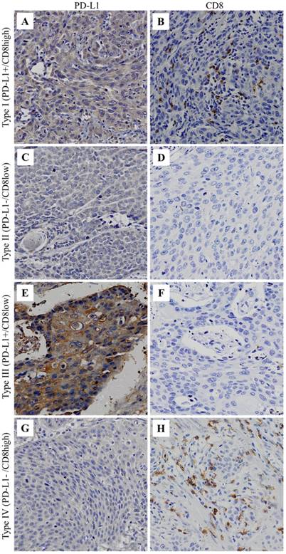
Type of TIM in ESCC tissues (×100). A and B, the same patient with type I TIM. C and D, the same patient with type II TIM. E and F, the same patient with type III TIM. G and H, the same patient with type IV TIM.

TIM classification based on PD-L1 expression and CD8+ T cell infiltration
The TIM of ESCC were classified into four types according to PD-L1 expression and CD8+ T cell infiltration (Figure 1). The distribution of the four types was 22.0% for type I, 26.0% for type II, 39.7% for type III and 12.3% for type IV. The associations between clinicopathological features and type of TIM were shown in Table 2. The results showed that only differentiation status was significantly associated with TIM (P=0.001).
Association between clinicopathological features and tumor immune microenvironment.
| Characteristics | PD-L1+, CD8 high | PD-L1-, CD8 low | PD-L1+, CD8 low | PD-L1-, CD8 high | P |
|---|---|---|---|---|---|
| Gender | |||||
| Male | 26(23.4%) | 25(22.5%) | 47(42.3%) | 13(11.7%) | 0.307 |
| Female | 6(17.1%) | 13(37.1%) | 11(31.4%) | 5(14.3%) | |
| Age | |||||
| ≤60 | 16(20.5%) | 21(26.9%) | 31(39.7%) | 10(12.8%) | 0.972 |
| >60 | 16(23.5%) | 17(25.0%) | 27(39.7%) | 8(11.8%) | |
| Tumor location | |||||
| Upper third | 4(14.3%) | 9(32.1%) | 12(42.9%) | 3(10.7%) | 0.428 |
| Middle third | 17(23.9%) | 21(29.6%) | 27(38.0%) | 6(8.5%) | |
| Lower third | 11(23.4%) | 8(17.0%) | 19(40.4%) | 9(19.1%) | |
| Tumor size (cm) | |||||
| ≤3 | 11(21.2%) | 17(32.7%) | 17(32.7%) | 7(13.5%) | 0.510 |
| 3<, ≤5 | 8(16.3%) | 13(26.5%) | 21(42.9%) | 7(14.3%) | |
| >5 | 13(29.5%) | 8(18.2%) | 19(43.2%) | 4(9.1%) | |
| Differentiation status | |||||
| Well | 11(14.9%) | 22(29.7%) | 31(41.9%) | 10(13.5%) | 0.001 |
| Moderately | 9(17.3%) | 11(21.2%) | 24(46.2%) | 8(15.4%) | |
| Poorly | 12(60.0%) | 5(25.0%) | 3(15.0%) | 0(0%) | |
| Tumor depth | |||||
| T1 | 4(25.0%) | 4(25.0%) | 4(25.0%) | 4(25.0%) | 0.259 |
| T2 | 7(14.6%) | 12(25.0%) | 24(50.0%) | 5(10.4%) | |
| T3 | 19(23.8%) | 22(27.5%) | 30(37.5%) | 9(11.3%) | |
| T4 | 2(100%) | 0(0%) | 0(0%) | 0(0%) | |
| Lymph node metastasis | |||||
| N0 | 16(19.8%) | 22(27.2%) | 31(38.3%) | 12(14.8) | 0.362 |
| N1 | 12(21.4%) | 15(26.8%) | 23(41.1%) | 6(10.7%) | |
| N2 | 0(0%) | 1(25.0%) | 3(75.0%) | 0(0%) | |
| N3 | 4(80.0%) | 0(0%) | 1(20.0%) | 0(0%) | |
| TNM stage | |||||
| Ⅰ | 5(16.1%) | 6(19.4%) | 12(38.7%) | 8(25.8%) | 0.153 |
| Ⅱ | 13(19.4%) | 21(31.3%) | 28(41.8%) | 5(7.5%) | |
| Ⅲ | 14(29.2%) | 11(22.9%) | 18(37.5%) | 5(10.4%) |
Univariate analysis of risk factors for overall survival of ESCC patients.
| Prognostic factors | β | HR (95% CI) | P value |
|---|---|---|---|
| Gender | -0.394 | 0.674(0.397-1.145) | 0.145 |
| Age | 0.044 | 1.045(0.688-1.588) | 0.835 |
| Tumor location | -0.065 | 0.937(0.689-1.258) | 0.667 |
| Tumor size | 0.073 | 1.076(0.830-1.394) | 0.580 |
| Differentiation status | 0.171 | 1.186(0.880-1.597) | 0.262 |
| Tumor depth | 0.547 | 1.727(1.224-2.438) | 0.002 |
| Lymph node metastasis | 0.439 | 1.551(1.181-2.036) | 0.002 |
| TNM stage | 0.518 | 1.678(1.242-2.267) | 0.001 |
| PD-L1 expression | 0.589 | 1.803(1.147-2.834) | 0.011 |
| CD8+ cell infiltration | 0.122 | 1.130(0.732-1.746) | 0.581 |
Multivariate analysis of risk factors for prognosis of ESCC patients.
| Prognostic factors | β | HR (95% CI) | P value |
|---|---|---|---|
| Tumor depth | 0.461 | 1.586(1.108-2.269) | 0.012 |
| Lymph node metastasis | 0.273 | 1.314(0.985-1.753) | 0.063 |
| PD-L1 expression | 0.496 | 1.643(1.038-2.601) | 0.034 |
Survival analysis
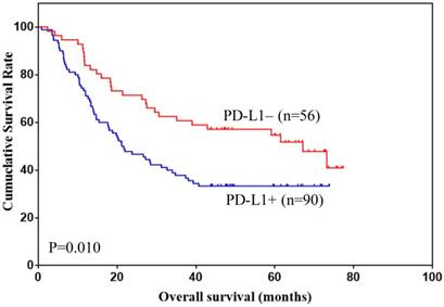
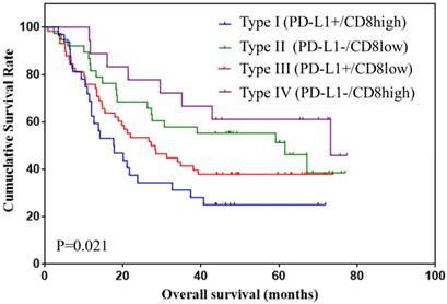
The 1, 3 and 5 years overall survival was 76.0%, 45.9% and 39.7%, respectively. The risk factors for the prognosis of ESCC patients were analyzed using univariate analysis (Table 3). The results showed that tumor depth, lymph node metastasis, TNM stage and PD-L1 expression were risk factors for the prognosis of ESCC patients (all P<0.05). However, only tumor depth and PD-L1 expression were independent prognostic risk factors (both P<0.05). The overall survival of ESCC patients according to PD-L1 expression was shown in Figure 2. The prognosis of patients with positive PD-L1 expression was significantly lower than that with negative PD-L1 expression. The overall survival of patients stratified by type of TIM were also analyzed and shown in Figure 3. The 5-year overall survival of Type I, II, III and IV was 24.7%, 40.8%, 32.3% and 50.2%, respectively.
Overall survival according to the PD-L1 expression on tumor cells.

Overall survival according to type of TIM.

Discussion
Although the associations between PD-L1 expression on tumor cells and prognosis of ESCC patients have been investigated in a series of studies, the findings were limited and controversial. Thus, the present study aims to investigate the prognostic value of PD-L1 expression and type of TIM in ESCC patients. We found that PD-L1 expression and CD8+ T cell infiltration was not associated with the clinicopathological features of ESCC. PD-L1 expression was an independent risk factor for the prognosis of ESCC. Immunotherapy may achieve promising outcomes in patients with type I TIM.
PD-L1 expression was studied limitedly in ESCC. Most reports evaluated the expression of PD-L1 by immunohistochemistry. Positive expression of PD-L1 was most commonly evaluated by the percentage of tumor cells with PD-L1 expression [21,22,28-30]. In the above mentioned five studies, positive rate of PD-L1 expression was 18.4%, 50.7%, 41.1%, 79.7% and 33.5%, respectively. Considering the positive rate of PD-L1 expression could not reflect the intensity of PD-L1 expression, IRS was employed in our present study. IRS ≥ 3 was considered to be PD-L1 positive expression. As a result, the positive rate of PD-L1 expression was 61.7% in our present study. The differences of positive rate of PD-L1 expression may attribute to the different antibody of PD-L1 during immunohistochemistry, sample size and definition of PD-L1 expression.
The associations between clinicopathological features and PD-L1 expression were inconsistent in the previous reports. The majority of studies showed that none of the clinicopathological features was associated with PD-L1 expression [21,22,30,31]. However, Jiang et al. and Ito et al. reported that PD-L1 expression was associated with deeper tumor depth and lymph node metastasis [29,32]. Interestingly, Chen et al. found that PD-L1 expression was correlated with better tumor differentiation, lower third tumor, N0 stage and earlier tumor stage [28]. In our present study, we also did not find any association between clinicopathological features and PD-L1 expression. Findings about the associations between PD-L1 expression and prognosis of ESCC patients in the previous reports were controversial [19,22,28,29,32-38]. The majority of investigations reported that PD-L1 expression was associated with unfavorable prognosis of ESCC patients [19,29,32-35,37,38]. However, a few studies reported the opposite results [22,28,36]. Recently, a meta-analysis, including 8 studies with a number of 1350 ESCC patients, indicated that PD-L1 expression was correlated with poorer overall survival of ESCC patients [39]. In our present study, we also found that PD-L1 expression was an independent risk factor for unfavorable overall survival of ESCC patients.
CD8+ T cell plays an important role in the anti-tumor immunity [40], and the associations between CD8+ T cell infiltration and clinicopathological features of ESCC were investigated widely. The majority of studies showed that none of the clinicopathological features was associated with CD8+ T cell infiltration in ESCC tissues [21,29,31,41]. However, two reports showed that CD8+ T cell infiltration was associated with male gender and elder patients, respectively [42,43]. Our present study observed a significant association between low density of CD8+T cell infiltration and well differentiation status [44]. It has been reported that CD8+ T cell infiltration was correlated with favorable prognosis of ESCC [42,45]. However, other studies did not find any association between CD8+ T cell infiltration and prognosis of ESCC patients [21,29,43,46]. In our present study, we also found that CD8+ T cell infiltration was not associated with the prognosis of ESCC. The inconsistent results in different studies may attribute to race, sample size, and especially the components of infiltrating immune cells. It was reported that the prognosis of ESCC was also associated with the infiltration of regulatory T cells, neutrophils, tumor associated macrophages, dendritic cells and monocytes, etc. [21,42,43,47]. This may influence the results about prognostic value of CD8+ T cell infiltration in ESCC in the previous reports.
Type of TIM was reported to be associated with the prognosis of gastric cancer [48,49]. Ma et al. demonstrated that PD-L1-/CD8+ gastric cancer had the best overall survival, and PD-L1+/CD8- gastric cancer had the worst overall survival [48]. Koh et al. also showed that PD-L1-/CD8 high gastric cancer had the best overall survival, but PD-L1-/CD8 low gastric cancer had the worst overall survival [49]. Up to date, prognostic value of TIM by co-assessment of PD-L1 expression and CD8+ T cell infiltration has not been investigated in ESCC yet. Only two study showed that PD-L1-/CD3 low subgroup or PD-L1+ /TIL low subgroup associated with worse DFS and OS in ESCC patients, respectively [31,35]. Recently, a study showed that the high intratumoral CD3 infiltration was correlated with favourable OS and DFS. Moreover, type classification based on intratumoral CD3 infiltration and PD-L1 expression on tumor cell was an independent prognostic factor for nasopharyngeal carcinoma patients. Nevertheless, peritumoral CD3 infiltration showed non prognostic value for OS and DFS[50]. These observations revealed the importance of local complex interaction microenvironment. In present study, for the first time, we investigated the value of TIM classification based on intratumoral CD8+ T cell infiltration and PD-L1 expression on tumor cell, and found that PD-L1-/CD8 high had the best overall survival, whereas PD-L1+/CD8 high had the worst overall survival. PD-L1 expression on tumor cells could favorably prognoses clinical outcome in nasopharyngeal carcinoma patients with pre-existing intratumor CD3 infiltrating lymphocytes[50]. It was reported that tumor regression after PD-L1/PD-1 blockade requires pre-existing CD8+ T cells [9]. In our study, we found that PD-L1+/CD8 high had the worst overall survival. From a treatment standpoint, this type of TIM maybe gain the maximum healing effect in potential immunotherapy of ESCC patients. Therefore, our findings indicated that type I (PD-L1+/CD8 high) ESCC could serve as candidate for anti-PD-1 or anti-PD-L1 immunotherapy.
There were some limitations in our present study. First, it was a retrospective study at a single institution, and the sample was relatively small. The findings in our study need further confirmation based on multicenter analysis with large sample size. Second, we did not analyze other tumor infiltrating immune cells in ESCC tissues. Thirdly, this study didn't investigate the relevance between the PD1 status on CD8+ T cells and clinical parameters. Fourthly, the disease free survival and disease specific survival of ESCC patients were not recorded and analyzed.
Conclusion
For the first time, the present study analyzed the associations between clinicopathological features and prognosis of ESCC patients and type of TIM based on co-assessment of PD-L1 expression and CD8+ T cell infiltration. We found that PD-L1 expression was not associated with clinicopathological features of ESCC. However, it was independently correlated with poor overall survival of ESCC. Further, we found that type of TIM was significantly associated with the prognosis of ESCC, which indicated that ESCC with type I TIM (PD-L1+/CD8 high) may achieve promising clinical outcomes through PD-L1/PD-1 blockade immunotherapy.
Abbreviations
PD-L1: programmed cell death ligand 1, ESCC: esophageal squamous cell carcinoma, TIM: tumor immune microenvironment.
Acknowledgements
This study was supported in part by grants from the National Natural Scientific Foundation of China [NO. 31570907, 81300301, 81572306, 81502403].
Authors' contributions
Qiao Wang, Fan Feng and Fei Wang contributed equally to this work. Hongwei Zhang and Fan Feng conceived and designed this study. Qiao Wang and Fei Wang carried on the immunohistochemistry staining. Qiao Wang wrote this manuscript. Gaozan Zheng, Fei Wang searched the literatures. Guanghui Xu, Zhen Liu, and Shushang Liu inputted the data. Fan Feng, Fei Wang, Qiao Wang, Guanghui Xu, Zhen Liu, Shushang Liu, Man Guo, Xiao Lian analyzed the data.
Availability of data and materials
All relevant data of the study were already included in the manuscript.
Ethics approval and consent
This study was approved by the Ethics Committee of Xijing Hospital. Written informed consent was obtained from the patient for publication of this manuscript and any accompanying images.
Competing Interests
The authors have declared that no competing interest exists.
References
1. Fitzmaurice C, Dicker D, Pain A, Hamavid H, Moradi-Lakeh M, MacIntyre MF, Allen C, Hansen G, Woodbrook R, Wolfe C, Hamadeh RR, Moore A, Werdecker A, Gessner BD, Te AB, McMahon B. et al. The Global Burden of Cancer 2013. JAMA ONCOL. 2015;1(4):505-527
2. Chen W, Zheng R, Zeng H, Zhang S, He J. Annual report on status of cancer in China, 2011. Chin J Cancer Res. 2015;27(1):2-12
3. McDermott DF, Atkins MB. PD-1 as a potential target in cancer therapy. Cancer Med. 2013;2(5):662-673
4. Ahmadzadeh M, Johnson LA, Heemskerk B, Wunderlich JR, Dudley ME, White DE, Rosenberg SA. Tumor antigen-specific CD8 T cells infiltrating the tumor express high levels of PD-1 and are functionally impaired. BLOOD. 2009;114(8):1537-1544
5. Jiang HB, Yang TJ, Lu P, Ma YJ. Gene expression profiling of gastric cancer. Eur Rev Med Pharmacol Sci. 2014;18(15):2109-2115
6. Hegde PS, Karanikas V, Evers S. The Where, the When, and the How of Immune Monitoring for Cancer Immunotherapies in the Era of Checkpoint Inhibition. CLIN CANCER RES. 2016;22(8):1865-1874
7. Herbst RS, Soria JC, Kowanetz M, Fine GD, Hamid O, Gordon MS, Sosman JA, McDermott DF, Powderly JD, Gettinger SN, Kohrt HE, Horn L, Lawrence DP, Rost S, Leabman M, Xiao Y. et al. Predictive correlates of response to the anti-PD-L1 antibody MPDL3280A in cancer patients. NATURE. 2014;515(7528):563-567
8. Tumeh PC, Harview CL, Yearley JH, Shintaku IP, Taylor EJ, Robert L, Chmielowski B, Spasic M, Henry G, Ciobanu V, West AN, Carmona M, Kivork C, Seja E, Cherry G, Gutierrez AJ. et al. PD-1 blockade induces responses by inhibiting adaptive immune resistance. NATURE. 2014;515(7528):568-571
9. Fehrenbacher L, Spira A, Ballinger M, Kowanetz M, Vansteenkiste J, Mazieres J, Park K, Smith D, Artal-Cortes A, Lewanski C, Braiteh F, Waterkamp D, He P, Zou W, Chen DS, Yi J. et al. Atezolizumab versus docetaxel for patients with previously treated non-small-cell lung cancer (POPLAR): a multicentre, open-label, phase 2 randomised controlled trial. LANCET. 2016;387(10030):1837-1846
10. Rosenberg JE, Hoffman-Censits J, Powles T, van der Heijden MS, Balar AV, Necchi A, Dawson N, O'Donnell PH, Balmanoukian A, Loriot Y, Srinivas S, Retz MM, Grivas P, Joseph RW, Galsky MD, Fleming MT. et al. Atezolizumab in patients with locally advanced and metastatic urothelial carcinoma who have progressed following treatment with platinum-based chemotherapy: a single-arm, multicentre, phase 2 trial. LANCET. 2016;387(10031):1909-1920
11. McDermott DF, Sosman JA, Sznol M, Massard C, Gordon MS, Hamid O, Powderly JD, Infante JR, Fasso M, Wang YV, Zou W, Hegde PS, Fine GD, Powles T. Atezolizumab, an Anti-Programmed Death-Ligand 1 Antibody, in Metastatic Renal Cell Carcinoma: Long-Term Safety, Clinical Activity, and Immune Correlates From a Phase Ia Study. J CLIN ONCOL. 2016;34(8):833-842
12. Stewart CL, Wilson L, Hamm A, Bartsch C, Boniface M, T D, SA P, SI J, H M, S Y, M K. Pembrolizumab (MK-3475) for patients (pts) with advanced esophageal carcinoma: Preliminary results from KEYNOTE-028. ASCO Annual Meeting. Chicago. 2015: Abstract 4010. 2017
13. T U, K M, H H, K Y, S H, S I, Kato K EA. Phase 2 study of Nivolumab (Anti-PD-1; ONO-4538) in Patients with Esophageal Cancer: Preliminary Report. The European Cancer Congress. Vienna, 2015: Abstract 2301. 2015
14. Taube JM, Anders RA, Young GD, Xu H, Sharma R, McMiller TL, Chen S, Klein AP, Pardoll DM, Topalian SL, Chen L. Colocalization of inflammatory response with B7-h1 expression in human melanocytic lesions supports an adaptive resistance mechanism of immune escape. SCI TRANSL MED. 2012;4(127):127r-137r
15. Wu P, Wu D, Li L, Chai Y, Huang J. PD-L1 and Survival in Solid Tumors: A Meta-Analysis. PLOS ONE. 2015;10(6):e131403
16. Taube JM, Klein A, Brahmer JR, Xu H, Pan X, Kim JH, Chen L, Pardoll DM, Topalian SL, Anders RA. Association of PD-1, PD-1 ligands, and other features of the tumor immune microenvironment with response to anti-PD-1 therapy. CLIN CANCER RES. 2014;20(19):5064-5074
17. Garon EB, Rizvi NA, Hui R, Leighl N, Balmanoukian AS, Eder JP, Patnaik A, Aggarwal C, Gubens M, Horn L, Carcereny E, Ahn MJ, Felip E, Lee JS, Hellmann MD, Hamid O. et al. Pembrolizumab for the treatment of non-small-cell lung cancer. N Engl J Med. 2015;372(21):2018-2028
18. Chen L, Deng H, Lu M, Xu B, Wang Q, Jiang J, Wu C. B7-H1 expression associates with tumor invasion and predicts patient's survival in human esophageal cancer. Int J Clin Exp Pathol. 2014;7(9):6015-6023
19. Chen MF, Chen PT, Chen WC, Lu MS, Lin PY, Lee KD. The role of PD-L1 in the radiation response and prognosis for esophageal squamous cell carcinoma related to IL-6 and T-cell immunosuppression. ONCOTARGET. 2016;7(7):7913-7924
20. Ohigashi Y, Sho M, Yamada Y, Tsurui Y, Hamada K, Ikeda N, Mizuno T, Yoriki R, Kashizuka H, Yane K, Tsushima F, Otsuki N, Yagita H, Azuma M, Nakajima Y. Clinical significance of programmed death-1 ligand-1 and programmed death-1 ligand-2 expression in human esophageal cancer. CLIN CANCER RES. 2005;11(8):2947-2953
21. Hatogai K, Kitano S, Fujii S, Kojima T, Daiko H, Nomura S, Yoshino T, Ohtsu A, Takiguchi Y, Doi T, Ochiai A. Comprehensive immunohistochemical analysis of tumor microenvironment immune status in esophageal squamous cell carcinoma. ONCOTARGET. 2016;7(30):47252-47264
22. Jiang D, Song Q, Wang H, Huang J, Wang H, Hou J, Li X, Xu Y, Sujie A, Zeng H, Tan L, Hou Y. Independent prognostic role of PD-L1expression in patients with esophageal squamous cell carcinoma. ONCOTARGET. 2017;8(5):8315-8329
23. Ma C, Patel K, Singhi AD, Ren B, Zhu B, Shaikh F, Sun W. Programmed Death-Ligand 1 Expression Is Common in Gastric Cancer Associated With Epstein-Barr Virus or Microsatellite Instability. AM J SURG PATHOL. 2016;40(11):1496-1506
24. Jinesh GG, Manyam GC, Mmeje CO, Baggerly KA, Kamat AM. Surface PD-L1, E-cadherin, CD24, and VEGFR2 as markers of epithelial cancer stem cells associated with rapid tumorigenesis. Sci Rep. 2017;7(1):9602
25. Critelli R, Milosa F, Faillaci F, Condello R, Turola E, Marzi L, Lei B, Dituri F, Andreani S, Sighinolfi P, Manni P, Maiorana A, Caporali C, di Benedetto F, Del BM, De Maria N. et al. Microenvironment inflammatory infiltrate drives growth speed and outcome of hepatocellular carcinoma: a prospective clinical study. CELL DEATH DIS. 2017;8(8):e3017
26. Xue J, Chen C, Qi M, Huang Y, Wang L, Gao Y, Dong H, Ling K. Type Igamma phosphatidylinositol phosphate kinase regulates PD-L1 expression by activating NF-kappaB. ONCOTARGET. 2017;8(26):42414-42427
27. Jiang L, Su X, Zhang T, Yin X, Zhang M, Fu H, Han H, Sun Y, Dong L, Qian J, Xu Y, Fu X, Gavine PR, Zhou Y, Tian K, Huang J. et al. PD-L1 expression and its relationship with oncogenic drivers in non-small cell lung cancer (NSCLC). ONCOTARGET. 2017;8(16):26845-26857
28. Chen K, Cheng G, Zhang F, Zhang N, Li D, Jin J, Wu J, Ying L, Mao W, Su D. Prognostic significance of programmed death-1 and programmed death-ligand 1 expression in patients with esophageal squamous cell carcinoma. ONCOTARGET. 2016;7(21):30772-30780
29. Jiang Y, Lo A, Wong A, Chen W, Wang Y, Lin L, Xu J. Prognostic significance of tumor-infiltrating immune cells and PD-L1 expression in esophageal squamous cell carcinoma. ONCOTARGET. 2017;8(18):30175-30189
30. Kim R, Keam B, Kwon D, Ock CY, Kim M, Kim TM, Kim HJ, Jeon YK, Park IK, Kang CH, Kim DW, Kim YT, Heo DS. Programmed death ligand-1 expression and its prognostic role in esophageal squamous cell carcinoma. World J Gastroenterol. 2016;22(37):8389-8397
31. Jesinghaus M, Steiger K, Slotta-Huspenina J, Drecoll E, Pfarr N, Meyer P, Konukiewitz B, Bettstetter M, Wieczorek K, Ott K, Feith M, Langer R, Weichert W, Specht K, Boxberg M. Increased intraepithelial CD3+ T-lymphocytes and high PD-L1 expression on tumor cells are associated with a favorable prognosis in esophageal squamous cell carcinoma and allow prognostic immunogenic subgrouping. ONCOTARGET. 2017;8(29):46756-46768
32. Ito S, Okano S, Morita M, Saeki H, Tsutsumi S, Tsukihara H, Nakashima Y, Ando K, Imamura Y, Ohgaki K, Oki E, Kitao H, Mimori K, Maehara Y. Expression of PD-L1 and HLA Class I in Esophageal Squamous Cell Carcinoma: Prognostic Factors for Patient Outcome. ANN SURG ONCOL. 2016;23(Suppl 4):508-515
33. Leng C, Li Y, Qin J, Ma J, Liu X, Cui Y, Sun H, Wang Z, Hua X, Yu Y, Li H, Zhang J, Zheng Y, Wang W, Zhu J, Wang Q. Relationship between expression of PD-L1 and PD-L2 on esophageal squamous cell carcinoma and the antitumor effects of CD8(+) T cells. ONCOL REP. 2016;35(2):699-708
34. Lim SH, Hong M, Ahn S, Choi YL, Kim KM, Oh D, Ahn YC, Jung SH, Ahn MJ, Park K, Zo JI, Shim YM, Sun JM. Changes in tumour expression of programmed death-ligand 1 after neoadjuvant concurrent chemoradiotherapy in patients with squamous oesophageal cancer. EUR J CANCER. 2016;52:1-9
35. Yagi T, Baba Y, Ishimoto T, Iwatsuki M, Miyamoto Y, Yoshida N, Watanabe M, Baba H. PD-L1 Expression, Tumor-infiltrating Lymphocytes, and Clinical Outcome in Patients With Surgically Resected Esophageal Cancer. ANN SURG. 2017
36. Wakita A, Motoyama S, Nanjo H, Sato Y, Yoshino K, Sasaki T, Kawakita Y, Liu J, Imai K, Saito H, Minamiya Y. PD-L1 Expression Is a Prognostic Factor in Patients with Thoracic Esophageal Cancer Treated Without Adjuvant Chemotherapy. ANTICANCER RES. 2017;37(3):1433-1441
37. Tsutsumi S, Saeki H, Nakashima Y, Ito S, Oki E, Morita M, Oda Y, Okano S, Maehara Y. Programmed death-ligand 1 expression at tumor invasive front is associated with epithelial-mesenchymal transition and poor prognosis in esophageal squamous cell carcinoma. CANCER SCI. 2017;108(6):1119-1127
38. Momose K, Yamasaki M, Tanaka K, Miyazaki Y, Makino T, Takahashi T, Kurokawa Y, Nakajima K, Takiguchi S, Mori M, Doki Y. MLH1 expression predicts the response to preoperative therapy and is associated with PD-L1 expression in esophageal cancer. ONCOL LETT. 2017;14(1):958-964
39. Qu HX, Zhao LP, Zhan SH, Geng CX, Xu L, Xin YN, Jiang XJ. Clinicopathological and prognostic significance of programmed cell death ligand 1 (PD-L1) expression in patients with esophageal squamous cell carcinoma: a meta-analysis. J THORAC DIS. 2016;8(11):3197-3204
40. Williams MA, Bevan MJ. Effector and memory CTL differentiation. ANNU REV IMMUNOL. 2007;25:171-192
41. Chen K, Zhu Z, Zhang N, Cheng G, Zhang F, Jin J, Wu J, Ying L, Mao W, Su D. Tumor-Infiltrating CD4+ Lymphocytes Predict a Favorable Survival in Patients with Operable Esophageal Squamous Cell Carcinoma. Med Sci Monit. 2017;23:4619-4632
42. Cho Y, Miyamoto M, Kato K, Fukunaga A, Shichinohe T, Kawarada Y, Hida Y, Oshikiri T, Kurokawa T, Suzuoki M, Nakakubo Y, Hiraoka K, Murakami S, Shinohara T, Itoh T, Okushiba S. et al. CD4+ and CD8+ T cells cooperate to improve prognosis of patients with esophageal squamous cell carcinoma. CANCER RES. 2003;63(7):1555-1559
43. Wang J, Jia Y, Wang N, Zhang X, Tan B, Zhang G, Cheng Y. The clinical significance of tumor-infiltrating neutrophils and neutrophil-to-CD8+ lymphocyte ratio in patients with resectable esophageal squamous cell carcinoma. J TRANSL MED. 2014;12:7
44. Rizvi NA, Hellmann MD, Snyder A, Kvistborg P, Makarov V, Havel JJ, Lee W, Yuan J, Wong P, Ho TS, Miller ML, Rekhtman N, Moreira AL, Ibrahim F, Bruggeman C, Gasmi B. et al. Cancer immunology. Mutational landscape determines sensitivity to PD-1 blockade in non-small cell lung cancer. SCIENCE. 2015;348(6230):124-128
45. Schumacher K, Haensch W, Roefzaad C, Schlag PM. Prognostic significance of activated CD8(+) T cell infiltrations within esophageal carcinomas. CANCER RES. 2001;61(10):3932-3936
46. Zingg U, Montani M, Frey DM, Dirnhofer S, Esterman AJ, Went P, Oertli D. Tumour-infiltrating lymphocytes and survival in patients with adenocarcinoma of the oesophagus. Eur J Surg Oncol. 2010;36(7):670-677
47. Zhu Y, Li M, Mu D, Kong L, Zhang J, Zhao F, Li Z, Liu X, Bo C, Yu J. CD8+/FOXP3+ ratio and PD-L1 expression associated with survival in pT3N0M0 stage esophageal squamous cell cancer. ONCOTARGET. 2016;7(44):71455-71465
48. Ma J, Li J, Hao Y, Nie Y, Li Z, Qian M, Liang Q, Yu J, Zeng M, Wu K. Differentiated tumor immune microenvironment of Epstein-Barr virus-associated and negative gastric cancer: implication in prognosis and immunotherapy. ONCOTARGET. 2017;8(40):67094-67103
49. Koh J, Ock CY, Kim JW, Nam SK, Kwak Y, Yun S, Ahn SH, Park DJ, Kim HH, Kim WH, Lee HS. Clinicopathologic implications of immune classification by PD-L1 expression and CD8-positive tumor-infiltrating lymphocytes in stage II and III gastric cancer patients. ONCOTARGET. 2017;8(16):26356-26367
50. Zhu Q, Cai MY, Chen CL, Hu H, Lin HX, Li M, Weng DS, Zhao JJ, Guo L, Xia JC. Tumor cells PD-L1 expression as a favorable prognosis factor in nasopharyngeal carcinoma patients with pre-existing intratumor-infiltrating lymphocytes. ONCOIMMUNOLOGY. 2017;6(5):e1312240
Author contact
![]() Corresponding authors: Hongwei Zhang, Ph.D, Division of Digestive Surgery, Xijing Hospital of Digestive Diseases, Fourth Military Medical University, 127 West Changle Road, 710032, Xi'an, Shaanxi, China. Tel: +86-029-84771531; Fax: +86-029-84771531; Email: zhanghwfmmucom. And Fan Feng, Ph.D, Division of Digestive Surgery, Xijing Hospital of Digestive Diseases, Fourth Military Medical University, 127 West Changle Road, 710032, Xi'an, Shaanxi, China. Tel: +86-029-84771531; Fax: +86-029-84771531; Email: surgeonfengfancom.
Corresponding authors: Hongwei Zhang, Ph.D, Division of Digestive Surgery, Xijing Hospital of Digestive Diseases, Fourth Military Medical University, 127 West Changle Road, 710032, Xi'an, Shaanxi, China. Tel: +86-029-84771531; Fax: +86-029-84771531; Email: zhanghwfmmucom. And Fan Feng, Ph.D, Division of Digestive Surgery, Xijing Hospital of Digestive Diseases, Fourth Military Medical University, 127 West Changle Road, 710032, Xi'an, Shaanxi, China. Tel: +86-029-84771531; Fax: +86-029-84771531; Email: surgeonfengfancom.

 Global reach, higher impact