Impact Factor ISSN: 1837-9664




J Cancer 2026; 17(6):1117-1124. doi:10.7150/jca.130032 This issue Cite
Research Paper
Association between obesity, sex, medical comorbidities, and survival in cancer patients treated with immune checkpoint inhibitors
Mingjia Li1*  , Daniel J. Spakowicz1,2*, Songzhu Zhao3*, Hyunwoo Kwon2,4, Kenneth Chian8, Adam Khorasanchi1, Yizhen Guo7, Lai Wei3, Lingbin Meng1,2, Yuanquan Yang1,2, Asrar Alahmadi1, Regan M. Memmott1, Jacob Kaufman1,2, Kai He1,2, Peter G. Shields1,2, Thomas A. Mace6, Mitch A. Phelps7, Christopher C. Coss7, Zihai Li1,2, Kari L. Kendra1, David P. Carbone1,2, Gregory A. Otterson1, Carolyn J. Presley1,2#, Dwight H. Owen1,2#
1. Division of Medical Oncology, Department of Internal Medicine, The Ohio State University, Columbus, USA.
2. Pelotonia Institute for Immuno-Oncology, The Ohio State University, Columbus, USA.
3. Department of Biomedical Informatics, Center for Biostatistics, The Ohio State University, Columbus, USA.
4. Department of Internal Medicine, The Ohio State University, Columbus, USA.
5. College of Medicine Medical Scientist Training Program, The Ohio State University, Columbus, USA.
6. Division of Gastroenterology Hepatology and Nutrition, Department of Internal Medicine, The Ohio State University, Columbus, USA.
7. College of Pharmacy, The Ohio State University, Columbus, USA.
8. College of Medicine, The Ohio State University, Columbus, USA.
* Denotes co-first authorship
# Denotes co-senior authorship.
Received 2025-12-16; Accepted 2026-4-20; Published 2026-6-10
Abstract
Background: Obesity's impact on cancer treatment outcomes is poorly understood, especially in the context of immuno-oncology. This study explores how obesity and medical comorbidities are associated with overall survival in cancer patients receiving immune checkpoint inhibitors (ICIs). Additionally, considering the influence of sex on body composition in obesity, this study examines the relationship between sex, obesity, medical comorbidities, and survival.
Methods: This cohort study involved 688 patients with metastatic cancer received ICIs as first- or second-line therapy. Obesity was assessed using body mass index (BMI). Cox proportional hazard models and Kaplan-Meier survival analysis were used to examine associations between predictors and overall survival.
Results: Patients with higher BMI had longer overall survival, and hazard ratio (HR) for death was 0.83 (95% CI 0.73-0.95) for every 10 units increased in BMI. Additionally, patients belonged to the highest BMI group (≥ 40) had the lowest risk of death when comparing to patients with BMI 18.5 to < 30 with HR 0.58 (95% CI 0.37-0.90). In subgroup analysis, a significant association between high BMI and decreased HR for death was predominantly observed in the male cohort. In multivariate analysis, the prognostic value of BMI remained significant after adjusting for performance status, line of therapy, age-adjusted medical comorbidities, and cancer type.
Conclusions: Obesity was associated with decreased mortality risk for cancer patients who had received ICIs. There could be a sex-dependent association between survival benefit and obesity.
1. Introduction
Obesity is one of the most prevalent comorbidities [1]. According to the World Health Organization (WHO), 13% of people aged 18 or greater are obese worldwide [2]. In the United States, the Center for Disease Control estimated 42.4% of adults were obese [3]. Obesity has important implications in cancer care given its pivotal role in oncogenesis [4, 5]. Conservatively, 10-20% of cancers are contributed to obesity [6, 7].
The effects of obesity on health are complex. Obesity has profound influences on anti-cancer immunity, and has been linked with higher risk of developing cancer due to dysregulation on immune surveillance [8]. Earlier studies have demonstrated increased inflammatory cytokines such as tumor necrosis factor α, interferon γ, interleukin (IL) -1, and IL-6 in obese cancer patients [9, 10]. Aberrant production, distribution, and function of natural killer (NK) cells, T-lymphocytes, and antigen-presenting cells have been associated with obesity [11]. Furthermore, multiple studies have demonstrated upregulations in signal transducer and activator of transcription 3 (STAT3) and programmed cell death protein 1 (PD-1) in obesity, which is thought to occur via increased leptin levels [12-14]. In addition to altered immunity, obesity reflects underlying nutritional status and influences treatment outcome by affecting drug metabolism [15-17].
Prior to the wide adaptations of immune checkpoint inhibitors (ICIs), obesity was associated with increased mortality in all cancers [18, 19]. This negative association has been revisited in the era of ICI-based treatments. Multiple retrospective studies have reported higher response rates and longer survival in obese cancer patients treated with ICIs [20-23]. In addition, this observed “obesity paradox” was more profound in male population [24, 25]. Although the exact mechanisms pertaining to obesity paradox have remained unclear, one potential mechanism proposes is the exhausted PD-1+ CD8+ T cells within obese immunosuppressive milieu have been more responsive to ICI. Furthermore, androgen-driven mechanism of CD8+ T cell exhaustion and improved PD-1 targeted therapy upon its blockade may contribute to increased ICI efficacy in obese males with marked alterations in androgen metabolism [26-30].
In this study, we investigated the relationships between obesity, sex, and clinical outcomes for patients with metastatic cancer treated with ICIs.
2. Methods
2.1 Study Design and Sample
We conducted a retrospective cohort study of 688 patients with metastatic cancer treated with immune checkpoint inhibitors. This study was reviewed and approved by the institutional review board (IRB 2016C0070). All patients received at least 1 dose of an immune checkpoint inhibitor as either first- or second-line therapy between 2011 and 2018 and remained under follow up until July 2020.
Clinical variables such as patients' demographics, tumor characteristics, and survival outcomes were collected via chart review. BMI was collected at baseline prior to the start of ICIs. BMI was defined as weight in kilograms divided by square height in meters (BMI = kg/m2). Charlson Comorbidity Index (CCI) was abstracted through query of ICD-10 codes for each patient [31]. Comorbidity data was manually extracted via chart review for 13 out of 688 patients in the all-cancer cohort who had missing ICD-10 data. A Modified CCI score excluding points assigned for cancer diagnosis was used for this study (Supplemental Table 1). Obesity was defined according to the WHO classification, which categorizes BMI ranges to determine obesity levels: obesity (class 1 30.0-34.9 and class 2 35.0-39.9), and extreme obesity (class 3) ≥ 40.
2.2 Statistical analysis
Patient characteristics were summarized using descriptive statistics including means (standard deviations) and median (interquartile ranges) for continuous variables and frequencies (percentages) for categorical variables. Pearson correlation was used to determine the correlation between BMI and modified CCI. The primary outcome of this study was overall survival, which was calculated from the date of ICI initiation until date of death from any cause or censored at loss to follow-up. Natural Cubic Spline was used to examine the potential non-linear association between BMI and overall survival with BMI = 30 chosen as the reference point. We have merged the groups of patients with normal weight (BMI 18.5-24.9) and overweight (BMI 25.0-29.9) into one category, and similarly combined Obese Class I (BMI 30.0-34.9) with Obese Class II (BMI 35.0-39.9) into another, based on our analysis using the natural cubic spline method. Cox proportional hazard models were first used to assess univariate associations between potential predictors for OS. After assessing collinearity, variables with a p-value < 0.05 (excluding BMI) were then entered into multivariable models for OS. Variables were removed sequentially from the multivariable models via backward selection. The associations of BMI with OS were modeled by Cox proportional hazards regression, controlling for the predictors retained after backward selection (modified CCI, line of therapy, and ECOG) and for cancer type, which was included based on clinical relevance. Adjusted hazard ratios (AHRs) and 95% CIs were displayed using forest plots. Patient characteristics were compared between males and females using two-sample t-test or Kruskal-Wallis test for continuous variables and chi square test for categorical variables. Heterogeneity by sex was evaluated by inserting an interaction term in the model (BMI*Sex). Survival curves were plotted using the Kaplan-Meier method and log-rank p-value was reported. R statistical software (version 4.2.0) was used to plot natural cubic splines and SAS (9.4) was used for the rest of the analysis.
3. Results
In our study, a total of 688 patients with metastatic cancers treated with first- or second-line ICIs were included. The mean BMI was 28.8 with standard deviation (SD) of 7.1. The median BMI was 28.0 with interquartile range (IQR) of 23.9 and 32.4. The study cohort included 285 (41%) females and 403 (59%) males, while 649 (94%) were white and 31 (5%) were black. The mean and median ages were 62 (with SD of 12.6) years and 62 (with IQR 54.3 and 70.3) years, respectively. A total of 360 patients (52%) received ICIs as first-line therapy and 328 (48%) received ICIs as second-line therapy. The majority of patients (569, 83%) had Eastern Cooperative Oncology Group performance status (ECOG) of 0 or 1 (Table 1).
Patient demographic and clinical characteristics with corresponding univariate hazard ratios (HRs) for overall survival. Cox proportional hazards models were used to examine univariate associations between risk factors and overall survival.
| Mean (SD) | HR (95% CI) | p-value | |
|---|---|---|---|
| Age | 61.9 (12.6) | 1.02 (1.01-1.02) | < 0.001 |
| BMI | 28.8 (7.1) | 0.83* (0.73-0.95) | 0.007 |
| Median (IQR) | |||
| CCI | 3 [1, 4] | 1.12 (1.08-1.17) | < 0.001 |
| N (percent) | |||
| BMI Groups | 0.038 | ||
| 18.5 to <30 | 413 (60%) | Reference | |
| <18.5 | 25 (3.6%) | 1.40 (0.89-2.20) | |
| 30 to <40 | 210 (30.5%) | 1.00 (0.82-1.21) | |
| ≥40 | 40 (5.8%) | 0.58 (0.37, 0.90) | |
| Cancer Type | < 0.001 | ||
| NSCLC | 157 (22.8%) | Reference | |
| Melanoma | 277 (40.3%) | 0.52 (0.41-0.66) | |
| Renal Cell Ca | 67 (9.7%) | 0.81 (0.58-1.12) | |
| Other | 187 (27.2%) | 0.81 (0.64-1.03) | |
| Line of Therapy | < 0.001 | ||
| First Line | 360 (52.3%) | Reference | |
| Second Line | 328 (47.7%) | 1.98 (1.65-2.38) | |
| ECOG | < 0.001 | ||
| 0 | 286 (41.6%) | Reference | |
| 1 | 283 (41.1%) | 1.91 (1.55-2.34) | |
| ≥2 | 107 (15.6%) | 3.18 (2.46-4.11) | |
| Unknown | 12 (1.7%) | ||
| Race | 0.426 | ||
| White | 649 (94.3%) | Reference | |
| Black | 31 (4.5%) | 1.30 (0.86-1.98) | |
| Other | 8 (1.2%) | 0.84 (0.35-2.03) | |
| Sex | 0.673 | ||
| Female | 285 (41.4%) | Reference | |
| Male | 403 (58.6%) | 0.96 (0.80-1.15) | |
| Smoking status | 0.007 | ||
| No | 273 (39.7%) | Ref | |
| Yes | 415 (60.3%) | 1.29 (1.07, 1.56) | |
| Diabetes status | 0.012 | ||
| No | 548 (79.7%) | Ref | |
| Yes | 140 (20.3%) | 1.32 (1.06, 1.63) | |
| Immunotherapy | 0.006 | ||
| Anti-PD-1 | 412 (59.9%) | Reference | |
| Anti-PD-L1 | 26 (3.8%) | 1.73 (1.12-2.67) | |
| Anti-CTLA-4 | 156 (22.7%) | 0.79 (0.63-0.99) | |
| Anti-PD1+CTLA-4 | 68 (9.9%) | 0.89 (0.65-1.20) | |
| Other | 26 (3.8%) | 0.66 (0.40-1.09) |
Significant p-values < 0.05 are bolded. *Hazard ratio is reported per 10-unit increase in BMI. BMI denotes body mass index; CCI, modified Charlson Comorbidity Index; CTLA-4, cytotoxic T-lymphocyte antigen-4; ECOG, Eastern Cooperative Oncology Group performance status; IQR, interquartile range; SD, standard deviation; PD-1, programmed cell death protein 1; PD-L1, programmed death-ligand 1.
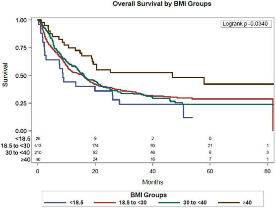
In this cohort, higher BMI was associated with longer OS. In univariate analysis, for every 10 unit increased in BMI, the hazard ratio (HR) of death decreased by 17% [HR = 0.83, 95% confidence interval (CI) 0.73-0.95, p = 0.007]. In natural cubic spline analysis, BMI < 20 was associated with accelerated increase in HR for death, whereas BMI ≥ 40 was associated with greater decline in HR (Supplemental Figure 1). In analyses using categorical BMI, compared to patients with BMI 18.5 to < 30, patients with morbid obesity (BMI ≥ 40) had a significantly decreased risk in death (HR 0.58, 95% CI 0.37-0.90) (Figure 1).
Kaplan-Meier analysis of overall survival Categorized by BMI. This analysis illustrates that patients with extreme obesity (BMI ≥ 40) exhibit the longest overall survival. BMI denotes body mass index.
To assess for underlying comorbidities, we used modified CCI by excluding points assigned to cancer. The median modified CCI was 3, and IQR 1 to 4. In univariate analysis, higher modified CCI at baseline was associated with increased mortality, HR 1.12 with 95% CI 1.08-1.17 for every point increase in modified CCI. There was no significant correlation between BMI and modified CCI. Pearson correlation coefficient was 0.011 and 2-tailed p-value was 0.77. In analysis of individual CCI components, significant associations were found between BMI groups and chronic obstructive pulmonary disease (p-value = 0.0438) and diabetes mellitus (p-value < 0.001).
In addition to BMI and modified CCI index, cancer type, line of therapy, ECOG performance status, smoking status, diabetes mellitus, type of immunotherapy was associated with overall survival (Table 1).
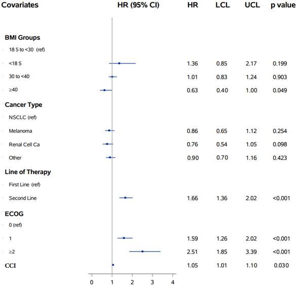
In multivariate analysis involving BMI group, modified CCI, cancer type, line of therapy, and ECOG performance status, BMI ≥ 40, line of therapy, ECOG, modified CCI index remained as significant predictor of death with p-value < 0.05 (Figure 2).
Multivariate analysis of BMI, cancer types, line of therapy, ECOG performance status, and modified CCI. This analysis reveals that a BMI ≥ 40, line of therapy, ECOG performance status, and the modified CCI are significant predictors of overall survival. BMI denotes body mass index; CCI, Charlson Comorbidity Index; ECOG, Eastern Cooperative Oncology Group; NSCLC, non-small cell lung cancer.
Sex Difference
We observed there is an association between BMI≥40 and overall survival in the male population, but not in the female population. In unadjusted model, for men, patients with BMI ≥40 had 65% lower hazard of death than those with BMI 18.5 to <30 (HR = 0.35, 95% CI 0.18-0.69). For women, there was no significant difference between BMI ≥40 and BMI 18.5 to <30 group. The interaction term for BMI ≥40 and sex was statistically significant (p = 0.016), indicating effect modification by sex for extreme obesity.
In multivariate analysis, after controlling for cancer type, ECOG performance status, line of therapy, and modified CCI, in males, BMI ≥40 remained a statistically significant predictor of OS (HR=0.44, 95% CI 0.22-0.91). Whereas for females, HR was 0.89 (0.49-1.63). Due to relatively low numbers of patients with BMI ≥40, the difference in HR between males and females was not significant, with the p-value for interaction term (sex*BMI) at 0.144 (Supplemental Table 2).
4. Discussion
The relationship between obesity, oncogenesis and cancer treatment outcome is complex. We hypothesized that immune dysfunction seen in obesity-related cancers contributed to longer overall survival for patients treated with ICI. In this study, we found that higher BMI was associated with improved overall survival in cancer patients treated with ICI. These findings are consistent with earlier studies linking higher BMI with survival [20, 21]. In addition, the greatest survival benefit was seen in patients with extreme obesity (BMI ≥40). Interestingly, in subgroup analysis, the association of BMI with longer survival was seen only in male patients and not in females.
To better assess the association between BMI and overall survival, we compiled a modified CCI to assess any potential underlying medical comorbidity that might have influenced treatment outcomes. The modified CCI in our study was strongly predictive of overall survival as previously reported [32]. However, we did not find a statistically significant correlation between increased BMI and modified CCI in our patients. The positive association between BMI and modified CCI were seen in multiple large population studies, and often not significant in smaller cohorts [33, 34]. In multivariate analysis after adjusting for ECOG performance status, cancer type, line of therapy, and modified CCI, BMI remained a statistically significant prognostic factor for overall survival.
Interestingly, a sex-biased association exists in our study between overall survival and increased BMI. In bivariate analysis, significant longer overall survival was observed only in male patients with extreme obesity (BMI ≥40). Furthermore, the difference in hazard ratios for death between males (HR 0.35, 95% CI 0.18-0.69) and females (HR 1.07, 95% CI 0.59-1.94) was substantial, with p=0.016 for the interaction term. In the multivariate analysis, extreme obesity remained a significant predictor of overall survival (OS) in males, but not in females, after adjustments for performance status, cancer type, line of therapy, and modified CCI. However, it is noteworthy that the difference in hazard ratios between males (HR 0.44, 95% CI 0.22-0.91) and females (HR 0.89, 95% CI 0.49-1.63) was not statistically significant, as indicated by a non-significant interaction between sex and BMI (p = 0.144), after adjusting for multiple potential confounders in our relatively small patient population with extreme obesity. To validate our findings, future studies with larger sample sizes are needed.
We hypothesize that the difference in male and female patients may be due to the differences in body composition between sex in our cohort. With the same BMI, males typically have higher visceral fat [35, 36]. Lower circulating androgen levels are associated with increased intra-abdominal fat in males [37]. In conjunction with recent published data, our clinical data lends support to androgen mediated T-cell exhaustion [26-28]. However, we do acknowledge our cohort had significantly fewer females, and only a low percentage of patients had extremely elevated BMI. Therefore, these analyses should be repeated in larger cohorts.
In addition, the positive association between high BMI and overall survival could contributed by the larger energy reserve in patients with obesity, which may offer greater ability to tolerate cancer treatment [38, 39]. It is well known that cancer associated cachexia is associated with poor outcome [40, 41]. BMI also reflects underlying body composition and affects pharmacokinetic/pharmacodynamic dosing of ICI with chemotherapies [17, 42, 43]. The effects of obesity on overall survival were beyond the scope of this project.
Taken together, although higher BMI was associated with improved overall survival, the effect size was modest and largely driven by patients with extreme obesity (BMI ≥40), indicating a nonlinear relationship. The nonlinear pattern suggests that the observed survival benefit may reflect underlying obesity-associated immune and metabolic states present in extreme obesity, rather than incremental differences in BMI alone. Importantly, these findings should not be interpreted as a recommendation for weight gain, as BMI likely reflects underlying host factors that influence response to immune checkpoint inhibition.
In our study, only the minority of patients (12%) had known PD-L1 status. Therefore, this variable was not included in the analysis due to the high level of missingness, which limits our ability to fully account for its potential impact on the overall survival analysis. However, existing clinical evidence regarding a direct association between BMI or obesity and tumor PD-L1 expression is inconsistent and appears to be cancer-specific and sex-dependent [44-46]. Notably, prior studies have shown that obesity or higher BMI may be independently associated with improved survival among patients with NSCLC receiving immunotherapy, including those with high PD-L1 expression [23, 47]. Larger, cancer-specific and sex-stratified cohorts are needed to further elucidate the relationship between obesity, PD-L1 expression, and immunotherapy outcomes.
This study has demonstrated the association between increased survival and high BMI after adjusting for medical comorbidities. There were several limitations in our study. First, this was a retrospective study and we could not establish any causal relationship. Second, although readily available, BMI could not measure body composition directly and we could not measure the distribution of adiposity [48]. Third, we were unable to effectively analyze ICI dosing and combination therapy with other cytotoxic chemotherapies due to the heterogeneity of our cohort. Finally, the lower number of female patients in our cohort could contribute to the lack of association between BMI and overall survival in females. Future studies consisting of a larger, more homogeneous cancer cohort are needed to further delineate some of these relationships.
5. Conclusion
Obesity was associated with increased overall survival for cancer patients who have received immune checkpoint inhibitors, independent of underlying medical comorbidities and performance status. Furthermore, the survival benefit related to obesity may vary depending on the patient's sex.
Abbreviations
BMI: body mass index
CCI: Charlson Comorbidity Index
CI: confidence interval
ECOG: Eastern Cooperative Oncology Group
HR: hazard ratio
ICI: immune checkpoint inhibitor
IL: interleukin
IQR: interquartile range
NSCLC: non-small cell lung cancer
OS: overall survival
RCC: renal cell carcinoma
STAT3: signal transducer and activator of transcription 3
PD-1: programmed cell death protein 1
PD-L1: programmed cell death-ligand 1
WHO: World Health Organization
Supplementary Material
Supplementary figures and tables.

Acknowledgements
Funding
This study was supported by the National Institutes of Health (P30CA016058, R01CA273924) and LUNGevity Foundation (Dwight H. Owen). Research support provided by the REDCap project and The Ohio State University Center for Clinical and Translational Science grant support (National Center for Advancing Translational Sciences, Grant UL1TR002733).
Data access
The deidentified datasets generated during and/or analyzed during the current study are available from the corresponding author upon reasonable request.
Ethical approval of studies
This study was approved by the Institutional Review Board at the Ohio State University (IRB 2016C0070).
Author contributions
Mingjia Li, Daniel J. Spakowicz, Carolyn J. Presley, and Dwight H. Owen contributed to the conception and design of the study. Mingjia Li, Daniel J. Spakowicz, Songzhu Zhao, Hyunwoo Kwon, Kenneth Chian, Adam Khorasanchi, Yizhen Guo, Lai Wei, Asrar Alahmadi, Regan Memmott, Jacob Kaufman, Kai He, Peter G. Shields, Kari L. Kendra, David P. Carbone, Gregory A. Otterson, Carolyn J. Presley, and Dwight H. Owen contributed to data acquisition. Songzhu Zhao and Lai Wei performed the statistical analysis. All authors contributed to data interpretation and critical review of the manuscript. Mingjia Li drafted the initial manuscript. All authors contributed to manuscript revision, reviewed and approved the final version.
Competing Interests
Carolyn J. Presley - Consulting or Advisory Role: OncLive, Regeneron.
Christopher Coss - Patent holder concerning IP discovered while employed by GTx INC. Patent holder and royalty recipient from Recursion Pharmaceuticals concerning IP surrounding methods of use for HDACi AR-42.
David P. Carbone - Consulting or Advisory Role: Merck, AstraZeneca, Bristol-Myers Squibb, EMD Serono, GlaxoSmithKline, Janssen, Genentech/Roche, Intellisphere, Lilly, Mirati Therapeutics, Johnson & Johnson/Janssen, Sanofi, Abbvie, Regeneron, PPD, Curio Science, Iovance Biotherapeutics, Jazz Pharmaceuticals, Merck KGaA, Novartis, Roche, InThought, OncLive/MJH Life Sciences, Pfizer, Arcus Biosciences, NCCN/AstraZeneca, MSD Oncology. Honoraria: AstraZeneca, Bristol-Myers Squibb/Ono Pharmaceutical.
Dwight H. Owen - Grants/funding/trial funding to institution from: Merck, Bristol-Myers Squibb, Genentech, Pfizer, Palobiofarma, Onc.AI, Nuvalent, Tango Therapeutics, Tubulis; honoraria from Chugai; and advisory to Roche (steering committee, uncompensated).
Gregory A. Otterson - Consult or Advisory Role: Novocure, OncLive/MJH Life Sciences. Research Funding: Genentech/Roche, Pfizer, Bristol-Myers Squibb, Novartis, Merck, AstraZeneca, Revolution Medicines, Array BioPharma, Apollomics.
He Kai - Consulting or Advisory Role: Perthera, Mirati Therapeutics, Bristol-Myers Squibb, Iovance Biotherapeutics, Geneplus, Lyell Immunopharma, and AstraZeneca. Research Funding: Bristol-Myers Squibb, Mirati Therapeutics, Adaptimmune, Genentech/Roche, GlaxoSmithKline, Amgen, Abbvie, Oncoc4.
Yuanquan Yang - Advisory Board: Exelixis, Eisai, Xencor. Consulting and Honoraria: The Whiteoak Group, AstraZeneca; Cardinal Health, Curio Science. Grant: NCI K12CA133250, Gateway for Cancer Research Foundation. Research support to institution: Incyte, Amgen, Novartis, Recordati Rare Diseases, Xencor, Taiho Oncology, AstraZeneca, AbbVie.
References
1. Ahima RS, Lazar MA. Physiology. The health risk of obesity-better metrics imperative. Science (New York, NY). 2013;341:856-8
2. Organization WH. Overweight and obesity. 2020.
3. Hales CM, Carroll MD, Fryar CD, Ogden CL. Prevalence of Obesity and Severe Obesity Among Adults: United States, 2017-2018. NCHS Data Brief. 2020:1-8
4. Steele CB, Thomas CC, Henley SJ, Massetti GM, Galuska DA, Agurs-Collins T. et al. Vital Signs: Trends in Incidence of Cancers Associated with Overweight and Obesity - United States, 2005-2014. MMWR Morb Mortal Wkly Rep. 2017;66:1052-8
5. De Pergola G, Silvestris F. Obesity as a Major Risk Factor for Cancer. Journal of Obesity. 2013;2013:291546
6. Wolin KY, Carson K, Colditz GA. Obesity and Cancer. The oncologist. 2010;15:556-65
7. Avgerinos KI, Spyrou N, Mantzoros CS, Dalamaga M. Obesity and cancer risk: Emerging biological mechanisms and perspectives. Metabolism. 2019;92:121-35
8. Woodall MJ, Neumann S, Campbell K, Pattison ST, Young SL. The Effects of Obesity on Anti-Cancer Immunity and Cancer Immunotherapy. Cancers (Basel). 2020 12
9. de Heredia FP, Gómez-Martínez S, Marcos A. Obesity, inflammation and the immune system. Proceedings of the Nutrition Society. 2012;71:332-8
10. Kern L, Mittenbühler MJ, Vesting AJ, Ostermann AL, Wunderlich CM, Wunderlich FT. Obesity-Induced TNFα and IL-6 Signaling: The Missing Link between Obesity and Inflammation-Driven Liver and Colorectal Cancers. Cancers. 2019;11:24
11. Kanneganti T-D, Dixit VD. Immunological complications of obesity. Nature Immunology. 2012;13:707-12
12. Bu LL, Yu GT, Wu L, Mao L, Deng WW, Liu JF. et al. STAT3 Induces Immunosuppression by Upregulating PD-1/PD-L1 in HNSCC. J Dent Res. 2017;96:1027-34
13. Li S-J, Wei X-H, Zhan X-M, He J-Y, Zeng Y-Q, Tian X-M. et al. Adipocyte-Derived Leptin Promotes PAI-1-Mediated Breast Cancer Metastasis in a STAT3/miR-34a Dependent Manner. Cancers. 2020;12:3864
14. Wang Z, Aguilar EG, Luna JI, Dunai C, Khuat LT, Le CT. et al. Paradoxical effects of obesity on T cell function during tumor progression and PD-1 checkpoint blockade. Nat Med. 2019;25:141-51
15. Hanley MJ, Abernethy DR, Greenblatt DJ. Effect of obesity on the pharmacokinetics of drugs in humans. Clin Pharmacokinet. 2010;49:71-87
16. Rush EC, Yan MR. Evolution not Revolution: Nutrition and Obesity. Nutrients. 2017;9:519
17. Centanni M, Moes DJAR, Trocóniz IF, Ciccolini J, van Hasselt JGC. Clinical Pharmacokinetics and Pharmacodynamics of Immune Checkpoint Inhibitors. Clinical Pharmacokinetics. 2019;58:835-57
18. Calle EE, Rodriguez C, Walker-Thurmond K, Thun MJ. Overweight, Obesity, and Mortality from Cancer in a Prospectively Studied Cohort of U.S. Adults. New England Journal of Medicine. 2003;348:1625-38
19. Basen-Engquist K, Chang M. Obesity and Cancer Risk: Recent Review and Evidence. Current Oncology Reports. 2011;13:71-6
20. Xu H, Cao D, He A, Ge W. The prognostic role of obesity is independent of sex in cancer patients treated with immune checkpoint inhibitors: A pooled analysis of 4090 cancer patients. International Immunopharmacology. 2019;74:105745
21. Nie RC, Chen GM, Wang Y, Yuan SQ, Zhou J, Duan JL. et al. Association Between Body Mass Index and Survival Outcomes In Patients Treated With Immune Checkpoint Inhibitors: Meta-analyses of Individual Patient Data. J Immunother. 2021;44:371-5
22. Hahn AW, Menk AV, Rivadeneira DB, Augustin RC, Xu M, Li J. et al. Obesity Is Associated with Altered Tumor Metabolism in Metastatic Melanoma. Clinical Cancer Research. 2022: OF1-OF11.
23. Kichenadasse G, Miners JO, Mangoni AA, Rowland A, Hopkins AM, Sorich MJ. Association Between Body Mass Index and Overall Survival With Immune Checkpoint Inhibitor Therapy for Advanced Non-Small Cell Lung Cancer. JAMA Oncology. 2020;6:512-8
24. Cortellini A, Bersanelli M, Buti S, Cannita K, Santini D, Perrone F. et al. A multicenter study of body mass index in cancer patients treated with anti-PD-1/PD-L1 immune checkpoint inhibitors: when overweight becomes favorable. J Immunother Cancer. 2019;7:57
25. McQuade JL, Daniel CR, Hess KR, Mak C, Wang DY, Rai RR. et al. Association of body-mass index and outcomes in patients with metastatic melanoma treated with targeted therapy, immunotherapy, or chemotherapy: a retrospective, multicohort analysis. Lancet Oncol. 2018;19:310-22
26. Kwon H, Schafer JM, Song NJ, Kaneko S, Li A, Xiao T. et al. Androgen conspires with the CD8(+) T cell exhaustion program and contributes to sex bias in cancer. Sci Immunol. 2022;7:eabq2630
27. Guan X, Polesso F, Wang C, Sehrawat A, Hawkins RM, Murray SE. et al. Androgen receptor activity in T cells limits checkpoint blockade efficacy. Nature. 2022;606:791-6
28. Yang C, Jin J, Yang Y, Sun H, Wu L, Shen M. et al. Androgen receptor-mediated CD8+ T cell stemness programs drive sex differences in antitumor immunity. Immunity. 2022;55:1268-83.e9
29. Pasquali R. Obesity and androgens: facts and perspectives. Fertil Steril. 2006;85:1319-40
30. Isidori AM, Caprio M, Strollo F, Moretti C, Frajese G, Isidori A. et al. Leptin and Androgens in Male Obesity: Evidence for Leptin Contribution to Reduced Androgen Levels*. The Journal of Clinical Endocrinology & Metabolism. 1999;84:3673-80
31. Charlson ME, Pompei P, Ales KL, MacKenzie CR. A new method of classifying prognostic comorbidity in longitudinal studies: Development and validation. Journal of Chronic Diseases. 1987;40:373-83
32. Johns AC, Wei L, Grogan M, Hoyd R, Bridges JFP, Patel SH. et al. Checkpoint inhibitor immunotherapy toxicity and overall survival among older adults with advanced cancer. J Geriatr Oncol. 2021;12:813-9
33. Song HJ, Hwang J, Pi S, Ahn S, Heo Y, Park S. et al. The impact of obesity and overweight on medical expenditures and disease incidence in Korea from 2002 to 2013. PloS one. 2018;13:e0197057
34. Afolabi HA, Zakariya Zb, Ahmed Shokri AB, Mohammad Hasim MNB, Vinayak R, Afolabi-Owolabi OT. et al. The relationship between obesity and other medical comorbidities. Obesity Medicine. 2020;17:100164
35. He X, Li Z, Tang X, Zhang L, Wang L, He Y. et al. Age- and sex-related differences in body composition in healthy subjects aged 18 to 82 years. Medicine (Baltimore). 2018;97:e11152
36. Bredella MA. Sex Differences in Body Composition. In: Mauvais-Jarvis F, editor. Sex and Gender Factors Affecting Metabolic Homeostasis, Diabetes and Obesity. Cham: Springer International Publishing. 2017 p. 9-27
37. Blouin K, Boivin A, Tchernof A. Androgens and body fat distribution. J Steroid Biochem Mol Biol. 2008;108:272-80
38. Demark-Wahnefried W, Platz EA, Ligibel JA, Blair CK, Courneya KS, Meyerhardt JA. et al. The Role of Obesity in Cancer Survival and Recurrence. Cancer Epidemiology, Biomarkers & Prevention. 2012;21:1244-59
39. Guo Y, Wei L, Patel SH, Lopez G, Grogan M, Li M. et al. Serum Albumin: Early Prognostic Marker of Benefit for Immune Checkpoint Inhibitor Monotherapy But Not Chemoimmunotherapy. Clinical lung cancer. 2022;23:345-55
40. Jain R, Coss C, Whooley P, Phelps M, Owen DH. The Role of Malnutrition and Muscle Wasting in Advanced Lung Cancer. Curr Oncol Rep. 2020;22:54
41. Roch B, Coffy A, Jean-Baptiste S, Palaysi E, Daures J-P, Pujol J-L. et al. Cachexia - sarcopenia as a determinant of disease control rate and survival in non-small lung cancer patients receiving immune-checkpoint inhibitors. Lung cancer (Amsterdam, Netherlands). 2020;143:19-26
42. Smit C, De Hoogd S, Brüggemann RJM, Knibbe CAJ. Obesity and drug pharmacology: a review of the influence of obesity on pharmacokinetic and pharmacodynamic parameters. Expert Opin Drug Metab Toxicol. 2018;14:275-85
43. Castillo AMM, Vu TT, Liva SG, Chen M, Xie Z, Thomas J. et al. Murine cancer cachexia models replicate elevated catabolic pembrolizumab clearance in humans. JCSM Rapid Commun. 2021;4:232-44
44. Berntsson J, Eberhard J, Nodin B, Leandersson K, Larsson AH, Jirström K. Pre-diagnostic anthropometry, sex, and risk of colorectal cancer according to tumor immune cell composition. Oncoimmunology. 2019;8:e1664275
45. Yugawa K, Itoh S, Iseda N, Kurihara T, Kitamura Y, Toshima T. et al. Obesity is a risk factor for intrahepatic cholangiocarcinoma progression associated with alterations of metabolic activity and immune status. Sci Rep. 2021;11:5845
46. Gałązka JK, Czeczelewski M, Kucharczyk T, Szklener K, Mańdziuk S. Obesity and lung cancer - is programmed death ligand-1 (PD-1L) expression a connection? Arch Med Sci. 2024;20:313-6
47. Cortellini A, Ricciuti B, Tiseo M, Bria E, Banna GL, Aerts JG. et al. Baseline BMI and BMI variation during first line pembrolizumab in NSCLC patients with a PD-L1 expression ≥ 50%: a multicenter study with external validation. J Immunother Cancer. 2020 8
48. Frankenfield DC, Rowe WA, Cooney RN, Smith JS, Becker D. Limits of body mass index to detect obesity and predict body composition. Nutrition. 2001;17:26-30
Author contact
Corresponding author: Mingjia Li, Email: Mingjia.LiEDU.