Impact Factor ISSN: 1837-9664




J Cancer 2026; 17(7):1393-1407. doi:10.7150/jca.128611 This issue Cite
Research Paper
Arteannuin B Induces Ferroptosis in Colorectal Cancer Cells via GDF15/HMGCS1/GPX4 Axis
Xiaozheng Ou1,2, Jinghong Xu2, Yuting Jin2, Yanqing Wang2, Rongmin Yu1,3, Jianhua Zhu1,3, Weijuan Huang1  , Liyan Song1,2
1. Biotechnological Institute of Chinese Materia Medica, Jinan University, Guangzhou, China.
2. Department of Pharmacology, College of Pharmacy, Jinan University, Guangzhou, China.
3. Department of Natural Medicinal Chemistry, College of Pharmacy, Jinan University, Guangzhou, China.
Received 2025-11-18; Accepted 2026-6-18; Published 2026-7-20
Abstract
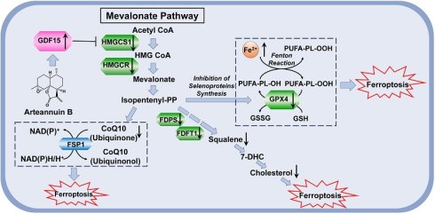
Purpose: Drug resistance in colorectal cancer (CRC) necessitates novel therapeutic strategies. This study investigated whether arteannuin B, a sesquiterpene lactone from Artemisia annua, induces ferroptosis in CRC cells and elucidated the underlying molecular mechanism.
Methods: Anti-CRC effects were assessed via MTT assays and xenograft models. Proteomics identified differentially expressed proteins. Arteannuin B-induced ferroptosis was confirmed by measuring reactive oxygen species (ROS), lipid peroxidation, Fe²⁺ content, glutathione peroxidase 4 (GPX4) expression, and mitochondrial morphology. Mevalonate pathway regulation was evaluated by western blotting, dual-luciferase assay, and quantification of squalene, coenzyme Q10 (CoQ10), and cholesterol. The role of growth differentiation factor 15 (GDF15) was validated using shRNA knockdown and overexpression DLD-1 cells in vitro and in vivo.
Results: Arteannuin B showed significant anti-colorectal cancer activity both in vitro and in vivo. The proteomic analysis demonstrated that arteannuin B affected the mevalonate pathway and ferroptosis in DLD-1 cells, and strongly upregulated the expression of GDF15. Arteannuin B increased ROS, lipid peroxidation, malondialdehyde, and iron while decreasing GPX4 expression and causing mitochondrial shrinkage. Arteannuin B inhibited mevalonate pathway enzymes, particularly 3-hydroxy-3-methylglutaryl-CoA synthase 1 (HMGCS1), reducing squalene, CoQ10, and cholesterol. The knockdown of GDF15 weakened the inhibitory effect of arteannuin B on the mevalonate pathway and GPX4, and reduced the sensitivity of CRC cells to arteannuin B both in vitro and in vivo.
Conclusion: Arteannuin B triggers ferroptosis-like cell death in CRC cells and suppresses xenograft growth, in association with inhibition of the mevalonate pathway. GDF15 contributes to arteannuin B-mediated suppression of HMGCS1 and GPX4 and to ferroptosis sensitivity.
Keywords: Arteannuin B, Colorectal cancer, Ferroptosis, Mevalonate pathway, Growth differentiation factor 15.
Introduction
Colorectal cancer is a type of cancer with a high incidence rate worldwide, and it is also a leading cause of cancer-related deaths [1]. Researchers predict that the burden of colorectal cancer will increase to 3.2 million new cases and 1.6 million deaths by 2040 [2]. Systemic chemotherapy remains the primary treatment for advanced disease; however, commonly used agents, including 5-fluorouracil (5-FU), oxaliplatin, irinotecan, and capecitabine, frequently lead to acquired resistance following prolonged or repeated administration [3]. Therefore, developing new therapeutic drugs is necessary and urgent. In recent years, targeted induction of ferroptosis has emerged as a promising strategy for colorectal cancer (CRC) treatment and may also offer new opportunities for diagnosis and prognosis.
Ferroptosis is a novel type of cell death mediated by iron ions and over-oxidized polyunsaturated fatty acids [4]. Depletion of intracellular glutathione (GSH) and the deletion/inactivation of glutathione peroxidase 4 (GPX4) are thought to be the classical pathways causing ferroptosis [5]. With gradually deepening research, the mevalonate pathway (MVP), a highly conserved cholesterol synthesis and lipid metabolism pathway in eukaryotic cells, had been confirmed to directly affect the process of ferroptosis. MVP is considered to protect cancer cells from ferroptosis through feedback regulation of cholesterol on coenzyme Q10 (CoQ10) and squalene [6] as well as the FSP1-CoQ10-NAD(P)H axis [7]. And its intermediates isopentenyl pyrophosphate (IPP) [8,9], farnesyl pyrophosphate (FPP) and geranylgeranyl pyrophosphate (GGPP) [10,11] stabilize GPX4, while 7-Dehydrocholesterol (7-DHC) shields lipids from peroxidation [12]. Therefore, the development of ferroptosis inducer and related drugs around MVP is becoming a valuable research field.
Growth differentiation factor 15 (GDF15), a member of the TGF-β superfamily, is involved in a variety of biological processes, including cell growth and proliferation, inflammation, and notably, the regulation of lipid metabolism [13]. GDF15 has been widely reported to be associated with ferroptosis, but the molecular mechanism is still unclear. It is reported that GDF15 is significantly upregulated during erastin-induced ferroptosis in HT-1080 cells [14] and has been concluded to be a protective gene in the prognosis of colorectal cancer patients [15]. Overexpression of GDF15 inhibited the proliferation and migration of clear cell renal cell carcinoma (ccRCC) and induced ferroptosis [16]. In this article, we discovered a potential association between GDF15 and MVP. The GDF15-MVP axis that induces ferroptosis has been validated through in vitro and in vivo experiments, and these results may provide novel valuable information for the development of ferroptosis-based therapies.
Artemisia annua L. is a traditional herbal medicine that has been used for centuries to treat malaria [17], fever [18] and to induce sedation [19]. Arteannuin B (CAS number: 50906-56-4) is a sesquiterpene lactone isolated from A. annua. Previous reports have revealed that arteannuin B has the effect of combating osteoporosis [20], anti-inflammation [21,22] as well as resisting SARS-CoV-2 [23,24]. Also, arteannuin B was found to show cytotoxicity to various human tumor cell lines in vitro [25]. In the preliminary research, we found that arteannuin B enhanced the effectiveness of cisplatin on non-small cell lung cancer [26]. But so far, the anti-colorectal cancer effect of arteannuin B and its potential mechanisms are not yet fully understood.
Recent research findings suggested that artemisinin compounds, such as artemisinin, dihydroartemisinin and artesunate, could exert anticancer activity by inducing cell ferroptosis [27]. In this study, arteannuin B was first found to induce ferroptosis in CRC cells. Through systematic research on the anti-colorectal cancer effect of arteannuin B in vitro and in vivo, we demonstrated that arteannuin B induced ferroptosis in CRC cells through the GDF15/HMGCS1/GPX4 axis. This molecular mechanism differed from the reported induction of ferroptosis by artemisinin compounds, providing a new perspective on the biological process of ferroptosis and the treatment of colorectal cancer.
Material and Methods
Materials
Ferrostatin-1 (Fer-1), 3-methyladenine (3-MA), benzyloxycarbonyl-Val-Ala-Asp (OMe)-fluoromethylketone (Z-VAD-FMK), and necrostatin-1 (Nec-1) were purchased from Selleck (Houston, USA). Anti-3-hydroxy-3-methylglutaryl-CoA reductase (HMGCR) antibody was purchased from ABclonal (Wuhan, China). Anti-HMGCS1 antibody, anti-GDF15 antibody, anti-FDFT1 antibody, anti-β-actin antibody and anti-GAPDH antibody were purchased from Proteintech (Wuhan, China). Anti-GPX4 antibody was purchased from Cell Signaling Technology (Danvers, USA). Radioimmunoprecipitation assay (RIPA) buffer, ROS detection kit, malondialdehyde (MDA) detection kit, and bicinchoninic acid (BCA) assay kit were purchased from Beyotime (Shanghai, China). Arteannuin B micelles and arteannuin B microspheres (ABMs) were prepared by Livzon Pharmaceutical Group Inc. Arteannuin B was isolated as previously reported [26].
Cell lines
Human colorectal cancer cell lines (DLD-1, HCT 15, HCT 116, HT-29, SW480, SW620, RKO, COLO 320, and COLO 205), human normal intestinal epithelial cell line (HIEC-6), and human normal colon fibroblast cell line (CCD-18Co) were purchased from the National Collection of Authenticated Cell Cultures (Shanghai, China). The cells were cultured in Dulbecco's Modified Eagle's Medium (DMEM) or RPMI 1640, supplemented with 10% fetal bovine serum and penicillin-streptomycin (final concentration: 100 U/ml penicillin and 100 µg/ml streptomycin). All cells were incubated at 37 °C in a humidified atmosphere with 5% CO2.
Cytotoxicity assay
Arteannuin B (6.25 to 100 µM) was incubated with cells for 24, 48, and 72 h, respectively. After incubation with methylthiazolyldiphenyl tetrazolium bromide solution (5 mg/ml) in the medium for 4 h, dimethyl sulfoxide (DMSO) was used to dissolve formazan. Subsequently, the OD570nm value of each sample well was detected and the cell survival rate was calculated.
Inhibition of ferroptosis
Different inhibitors were applied to block the cell death caused by arteannuin B. After the pretreatment of Fer-1 (2 µM, 4 h), 3-MA (100 µM, 2 h), Z-VAD-FMK (2 μM, 2 h), and Nec-1 (5 µM, 2 h), DLD-1 cells were then treated with arteannuin B (15 µM) and different inhibitors for 24 h. Cell viability was detected through MTT assay.
ROS detection
ROS generation was detected by DCFH-DA staining. Briefly, DLD-1 cells were incubated with 5 μM DCFH-DA at 37 °C for 30 min after being treated with arteannuin B (7.5, 15, 30 µM) for 48 h. Quantitative analysis of intracellular ROS level was performed using flow cytometry according to the instructions of the ROS detection kit.
Measurement of lipid peroxidation
DLD-1 cells were seeded in 6-well plates and treated with arteannuin B (3.75, 7.5, 15 µM) for 48 h. The cells were then collected and re-suspended in 2 μM C11-BODIPY581/591 (Cayman Chemical, MI, USA) for 30 min at 37 °C. Subsequently, cells were measured by flow cytometry. Quantification of data was performed with FlowJo 10.8.1.
Detection of intracellular metabolites
DLD-1 cells were treated with arteannuin B for 48 h. Then, cells were lysed in RIPA buffer and the protein concentration was quantified by BCA assay kit. The intracellular MDA level was determined by the MDA detection kit. The intracellular iron level was determined by the iron assay kit (Sigma Aldrich, MO, USA). The content of squalene, cholesterol and CoQ10 in cell lysates was determined and quantified by squalene ELISA detection kit (Feiyue, Wuhan, China), total cholesterol assay kit (Solarbio, Shanghai, China) and CoQ10 ELISA detection kit (ZOKEYO, Wuhan, China).
Transmission electron microscopy
Transmission electron microscopy was used to observe the morphological changes of mitochondria in DLD-1 cells after administration of arteannuin B. Briefly, DLD-1 cells were treated with arteannuin B at a concentration of 15 µM for 48 h. Cells were collected and fixed in a fixation solution containing 2.5% glutaraldehyde. Subsequently, the samples were sent to Wuhan Servicebio Technology Co., Ltd (Wuhan, China) for dehydration, embedding, sectioning, and staining processes. Transmission electron microscope was used to capture the pictures.
Western blotting analysis
DLD-1 cells were treated with arteannuin B for 48 h and lysed in RIPA buffer. The cell lysates were centrifuged and protein concentration was measured using a BCA assay kit. Equal amounts of protein were separated by SDS-PAGE. The proteins were then transferred onto a polyvinylidene fluoride (PVDF) membrane and blocked with bovine serum albumin to prevent nonspecific binding. Next, the membrane was incubated with a primary antibody specific to the target protein, followed by an HRP-conjugated secondary antibody. Finally, the target protein was visualized using an enhanced chemiluminescence kit and chemiluminescence imaging system.
Real-time quantitative PCR analysis
Total RNA was isolated from cells or tissue samples using Trizol reagent (Beyotime, Shanghai, China) strictly following the manufacturer's instructions. During the extraction process, the volume of lysis buffer was carefully controlled, and only the upper aqueous phase was collected to avoid genomic DNA contamination caused by organic phase carry-over. The concentration and quality of the extracted total RNA were assessed using a NanoD2000C (Thermo, USA) to verify RNA purity.
Synthesis of cDNA was performed by RT-PCR from 1000 ng of total RNA using the Hifair® Ⅱ 1st Strand cDNA Synthesis SuperMix for qPCR (Yeasen, Shanghai, China), which contains a gDNA digester that specifically removes residual genomic DNA from RNA samples. To further ensure no genomic DNA amplification during the PCR step, a no-RT negative control was set for each group.
The sample mixture was prepared according to the TransStart® Top Green qPCR SuperMix (TransGen, Beijing, China) instructions. Then, RT-qPCR experiment was performed by LightCycler® 480 (Roche, Basel, Switzerland). All reactions were normalized to GAPDH level, and 2-ΔΔCt method was used for data analysis.
Proteomic analysis
Proteomic analysis was supported by Shanghai Applied Protein Technology Co. Ltd (Shanghai, China). Briefly, DLD-1 cells were incubated with or without 15 µM arteannuin B for 48 h. Cell lysates were then collected and subjected to tandem mass tag (TMT) relative quantitative proteomic analysis. The T/C value was defined as the difference in protein expression multiples between the arteannuin B treatment group and the normal control group. Proteins with a T/C value outside 0.83 to 1.2 and p < 0.05 were confirmed as significantly differentially expressed proteins (SDEPs). Bioinformatics analysis was performed on the identification results.
Survival Analysis
The Kaplan-Meier method was employed to construct overall survival curves for colorectal cancer patients using the GEPIA2 tool. The discrepancy in survivals was assessed through the utilization of the log-rank test, alongside the calculation of the HRs (hazard ratio with 95% confidence interval). The core data for GEPIA survival analysis were derived from two major international public databases: the Cancer Genome Atlas (TCGA) and the Genotype-Tissue Expression (GTEx).
Plasmid construction and cell transfection
GDF15 specific shRNA plasmids (shGDF15: 5'-AGACTCCAGATTCCGAGAGTT-3'), GDF15 overexpression plasmids, GPX4 overexpression plasmids, lentiviral plasmids, sterol regulatory element-binding protein 2 (SREBP2) plasmid and control plasmid, HMGCS1 promoter plasmid and control plasmid, and marine luciferase plasmid were purchased from Transheepbio (Shanghai, China).
HEK293T cells were incubated with lentiviral vectors, psPAX2 and pMD2G vectors, PEI (4 μg/ml) for virus production. After two days of normal culture of cells, the cells were then infected with the virus for 24 h. Puromycin (3 μg/ml) was used to select stably transduced cells for 1 week.
For the dual-luciferase reporter assay, DLD-1 cells were seeded at a density of 1×10⁶ cells per well in a six-well plate. When the cell density reached 70%, transient transfection was used to transfer the HMGCS1 promoter plasmid and promoter control plasmid. The detection of the binding of SREBP2 and HMGCS1 promoter before and after 15 μM arteannuin B was carried out using glow type dual luciferase reporter gene detection kit (Yeasen, Shanghai, China). A blank control group was set to subtract the background, and the expression fold was calculated as (firefly luciferase value of the experimental group/Renilla luciferase value)/(corresponding ratio of the control group).
In vivo anti-colorectal cancer experiment of arteannuin B
DLD-1 cells (5×10⁷ cells/mL) were subcutaneously injected (100 μL) into left axillary regions of male BALB/c nude mice (6-7 weeks old). When tumors reached approximately 70 mm³, mice were randomized into six groups receiving daily treatments for 21 days: model group (10 ml/kg normal saline), blank micelles group (blank micelles), arteannuin B micelles group (20 mg/kg arteannuin B micelles), vehicle group (1 ml/kg DMSO), arteannuin B group (20 mg/kg arteannuin B), and 5-FU group (20 mg/kg 5-FU). During the administration period, the tumor volume of mice in each group was detected every other day. Post-treatment, mice were euthanized for organ collection (liver, kidneys, spleen, heart) and histopathological analysis. Tumor tissues were cryopreserved (-80°C) and formalin-fixed. GPX4 expression in tumor specimens was assessed by immunohistochemistry (IHC).
In vivo experiments identifying GDF15 as a key target protein
DLD-1WT and DLD-1shGDF15 cells (2.0×10⁷ cells/mL) were subcutaneously injected (100 μL) into left axillary regions of male BALB/c nude mice (6-7 weeks old). Mice with tumors approximately 70 mm³ were randomized into six groups: DLD-1WT/Model group (10 ml/kg normal saline), DLD-1WT/Vehicle group (blank microspheres), DLD-1WT/ABMs group (140 mg/kg ABMs), DLD-1shGDF15/Model group (10 ml/kg normal saline), DLD-1shGDF15/Vehicle group (blank microspheres), DLD-1shGDF15/ABMs group (140 mg/kg ABMs), treated weekly for 4 weeks. During the administration period, the tumor volume of mice in each group was detected every other day. Post-treatment, mice were euthanized for organ collection (liver, kidneys, spleen, heart) and histopathological analysis. Tumor tissues were cryopreserved (-80°C) and formalin-fixed. GPX4 expression in tumor specimens was assessed by IHC, while organizational lysate was analyzed for MDA and cholesterol levels.
Statistical analysis
Statistical analysis was performed using SPSS (Version 25.0) and GraphPad Prism (Version 9.5.1). Graphs were established using GraphPad Prism (Version 9.5.1) and ImageJ (Version 1.5.4). Results were expressed as mean ± standard deviation (SD), n ≥ 3. Two-tailed Student's t-test was used to assess differences between two groups, and one-way analysis of variance (ANOVA) followed by Sidak's multiple comparisons test was used to count differences between multiple groups. Statistical significance was considered for p < 0.05.
Results
Anti-colorectal cancer effects of arteannuin B in vitro and in vivo
The results of the cytotoxicity experiment showed that arteannuin B exhibited potent anti-proliferative effects across a panel of 9 human colorectal cancer cell lines in a concentration- and time-dependent manner (Figure 1A), suggesting its broad-spectrum activity against CRC cells. After 72 h of treatment on DLD-1, COLO 205, COLO 320, HCT 15 and HT-29 cells, the IC50 values of arteannuin B were all less than 10 µM (Table S1). Based on the screening results, DLD-1 was identified as the most responsive cell line to arteannuin B and was therefore chosen as the primary model for subsequent mechanistic investigations.
Arteannuin B showed anti-colorectal cancer effects in vitro and in vivo. (A) Anti-proliferative effects of arteannuin B on nine human CRC cells at (a) 24 h, (b) 48 h, and (c) 72 h (n = 3). (B) Effect of arteannuin B on viability of CCD-18Co cells and HIEC-6 cells (n = 3). (C-D) Effect of arteannuin B on tumor volume and tumor weight of nude mice xenografted with DLD-1 cells (n = 6). (E) Pictures of tumor tissues. (F) Changes of the body weight of animals (n = 6). Data are presented as mean ± SD; *p < 0.05, **p < 0.01 vs. model group.
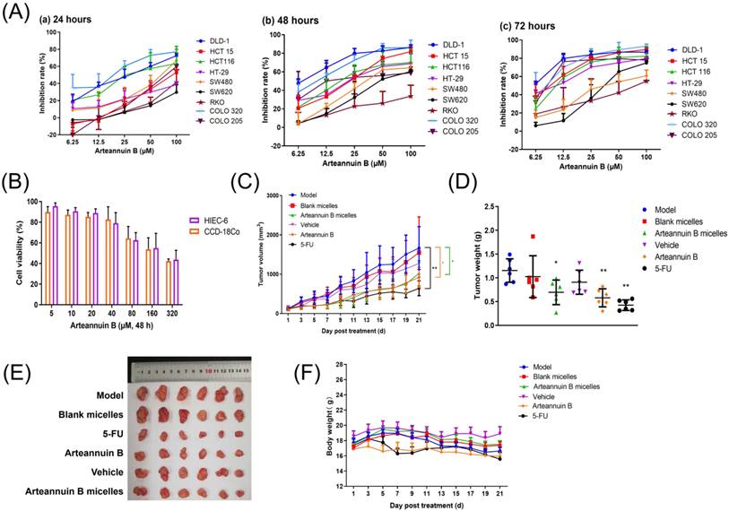
When arteannuin B was applied to human normal colon fibroblasts CCD-18Co and human normal intestinal epithelial cells HIEC-6 for 48 h, the IC50 values of arteannuin B were 165.15 ± 49.56 μM and 228.49 ± 72.94 μM, respectively (Figure 1B). Within the commonly used concentration range from 3.75 to 15 μM in this study, arteannuin B showed no significant effect on the cell viability of normal colon cells (the cell viability is greater than 85%), but selectively inhibited the proliferation of CRC cells.
The DLD-1 cell xenograft tumor model was established to detect the in vivo anti-tumor effect of arteannuin B. The experimental results demonstrated that 20 mg/kg arteannuin B significantly inhibited the growth of xenograft tumors (Figure 1C) with the inhibition rate of 49.71% (Figure 1D-E). After 21 d of arteannuin B treatment, the body weight of the animals in the treatment group showed no significant differences compared to the model group (Figure 1F), and the organ indices of mice remained unaffected (Table 1). These data showed that arteannuin B did not show obvious toxic effects under experimental conditions.
Organ indices of the nude mice xenografted with DLD-1 cells.
| Groups | Organ Indices (mg/g, x̄ ± SD, n = 6) | |||
|---|---|---|---|---|
| Heart | Liver | Spleen | Kidney | |
| Model | 5.80 ± 1.55 | 45.95 ± 1.86 | 4.28 ± 1.12 | 13.12 ± 3.43 |
| Blank micelles | 4.76 ± 1.62 | 58.23 ± 2.66 | 4.68 ± 1.59 | 14.98 ± 1.18 |
| Arteannuin B micelles | 6.06 ± 1.38 | 58.75 ± 4.81 | 6.44 ± 1.94 | 14.54 ± 1.39 |
| Vehicle | 5.84 ± 1.50 | 51.31 ± 2.04 | 4.76 ± 0.72 | 14.78 ± 1.46 |
| Arteannuin B | 5.98 ± 1.12 | 52.96 ± 3.74 | 4.08 ± 1.93 | 15.40 ± 1.67 |
| 5-FU | 5.50 ± 1.07 | 47.99 ± 4.92 | 5.23 ± 2.05 | 15.41 ± 1.26 |
Proteomic analysis of the anti-colorectal cancer effect of arteannuin B
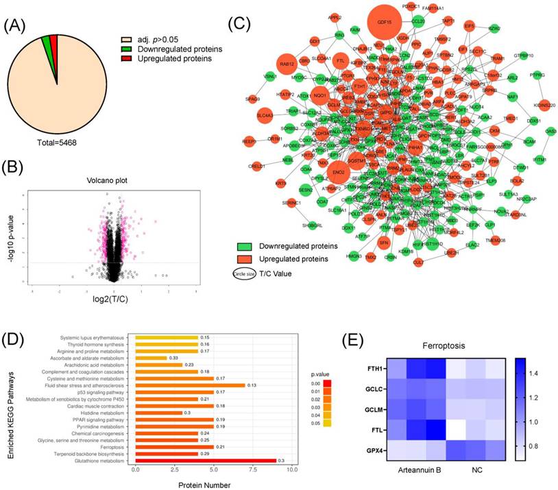
After treating DLD-1 cells with arteannuin B for 48 h, we extracted total protein for TMT-based quantitative proteomic analysis. A total of 5468 proteins were identified (Figure 2A). The T/C value was defined as the difference in protein expression multiples between the arteannuin B treatment group and the normal control group. When the T/C value was not within the range of 0.83 to 1.2 times and the p-value was less than 0.05, the protein was identified as being differentially expressed (Figure 2B). A total of 144 differentially upregulated proteins and 152 differentially downregulated proteins were identified. Among them, the most significantly upregulated protein is GDF15 (T/C=2.869102), and the most significantly downregulated protein is histone H1.3 (encoded by HIST1H1D) (T/C=0.527179) (Figure 2C). The results of Kyoto Encyclopedia of Genes and Genomes (KEGG) pathway enrichment analysis showed that SDEPs were mainly enriched in biological pathways related to glutathione metabolism, terpenoid backbone biosynthesis (mevalonate pathway), and ferroptosis pathway (Figure 2D). The proteins related to the ferroptosis pathway were involved in ferritin heavy chain (FTH1), glutamate-cysteine ligase catalytic subunit (GCLC), glutamate-cysteine ligase regulatory subunit (GCLM), ferritin light chain (FTL) and GPX4 (Figure 2E). Among them, FTH1, GCLC, GCLM, and FTL were observed to be significantly up-regulated, and these increases are likely to represent an active cellular defense aimed at limiting ferroptosis progression [28]. In contrast, the decreased expression of GPX4 is one of the most emblematic molecular events of ferroptosis.
Proteomic analysis of the anti-colorectal cancer effect of arteannuin B. (A) Overview of proteomics analysis results. (B) Volcano plot of SDEPs. (C) Visualization of SDEPs. (Green: downregulated proteins; Red: upregulated proteins; Size of the circle: T/C value). (D) KEGG analysis of SDEPs. (E) SDEPs enriched in the ferroptosis signaling pathway.
Arteannuin B induced ferroptosis in CRC cells
To determine whether arteannuin B induced ferroptosis in CRC cells, DLD-1 cells were pretreated with autophagy inhibitor (3-MA), apoptosis inhibitor (Z-VAD-FMK), necroptosis inhibitor (Nec-1), and ferroptosis inhibitor (Fer-1), respectively. The results showed that only the ferroptosis inhibitor could significantly relieve the inhibitory effect of arteannuin B on DLD-1 cells (Figure 3A).
Arteannuin B induced ferroptosis in CRC cells. (A) Influence of cell death inhibitors on the anti-proliferative effect of arteannuin B against DLD-1 cells. (B-D) Effects of arteannuin B on ROS, lipid peroxidation, MDA levels in DLD-1 cells. (E) Effects of arteannuin B on iron ions content in DLD-1 cells. (F-G) Western blotting images and quantitative analysis showed the effects of arteannuin B on GPX4 protein expression levels in DLD-1 cells. (H) Effects of arteannuin B on mRNA transcription level of GPX4 in DLD-1 cells. (I) Effects of arteannuin B on mitochondrial morphology of DLD-1 cells. In the control group, mitochondria are elongated rods with parallel cristae (upper panel). In the arteannuin B-treated group, mitochondria are smaller and rounded (yellow arrows), with a denser matrix (darker staining), discontinuous outer membrane, and loss of cristae (red arrows). Scale bar = 5 μm, 1 μm. Data are presented as mean ± SD, n = 3; &&p < 0.01 vs. arteannuin B group without inhibitor treatment; *p < 0.05, **p < 0.01, ***p < 0.001 vs. control group.
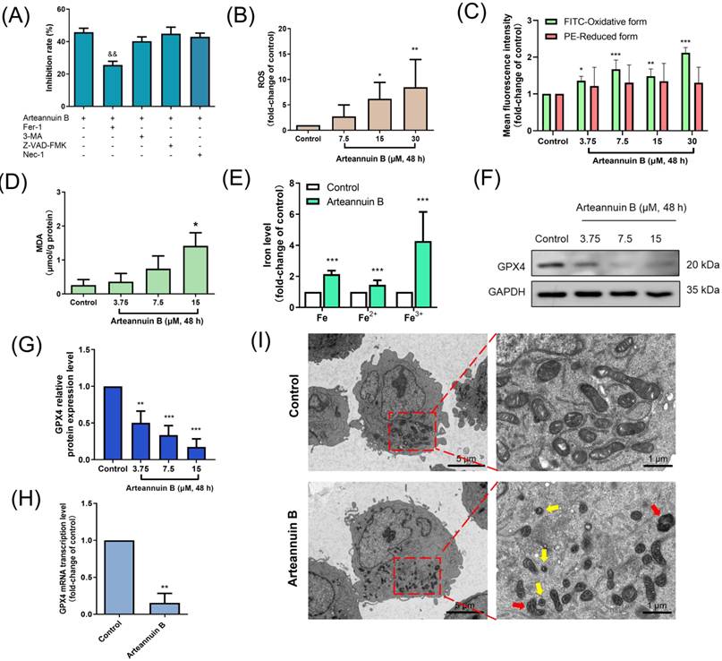
Out-of-control membrane lipid peroxidation is a critical step in inducing ferroptosis [29]. It was observed that arteannuin B significantly increased the content of ROS (Figure 3B) and the level of lipid peroxidation (Figure 3C) in DLD-1 cells. The level of MDA, a lipid peroxidation product, was also detected to increase after the treatment of arteannuin B (Figure 3D). These results indicated that arteannuin B disrupted the intracellular redox balance and induced lipid peroxidation in DLD-1 cells. In addition, arteannuin B leaded to an increase in iron ion level within CRC cells (Figure 3E). The iron accumulation caused by arteannuin B showed an increase in total intracellular iron content, rather than an increase only in Fe2+ content.
As mentioned earlier, GPX4 is a key protein in cells that resists ferroptosis. After the DLD-1 cells were treated with arteannuin B for 48 h, the protein expression level of GPX4 was significantly downregulated (Figure 3F-G), and the mRNA transcription level of GPX4 also showed a significant decrease (Figure 3H). In addition, arteannuin B also downregulated the protein expression level of GPX4 in the DLD-1 cells xenograft tumor tissues (Figure S1). These data indicated that GPX4 protein was significantly inhibited in DLD-1 cells after administration of arteannuin B.
The morphological changes of cell mitochondria are another important indicator of ferroptosis [30]. DLD-1 cells treated with 15 μM arteannuin B were examined for alterations in organelle morphology using transmission electron microscopy. As shown in Figure 3I, in normally cultured DLD-1 cells, mitochondria appeared as elongated rods with parallel-arranged cristae. After arteannuin B treatment, the mitochondrial volume was markedly reduced and became round (yellow arrows). The matrix showed high electron density (darkened), the outer membrane was discontinuous in places, and the cristae had disappeared (red arrows). Our results demonstrated that arteannuin B inhibited the proliferation of CRC cells by inducing ferroptosis.
GDF15 was identified as a key factor in arteannuin B-induced ferroptosis
Based on the data of proteomic analysis, GDF15 was found to be the most significantly upregulated protein in DLD-1 cells after the administration of arteannuin B (Figure 2C). In previous studies, a multitude of natural products and active ingredients of traditional Chinese medicine have been documented to elicit anti-neoplastic actions against colorectal carcinoma, potentially through the modulation of GDF15 and its associated signaling cascades [31,32]. Our subsequent research endeavors aimed to elucidate whether GDF15 represents a molecular target of arteannuin B in its anti-colorectal cancer activity, and to delineate the mechanistic role of GDF15 in arteannuin B-induced ferroptosis.
Consistent with the results of proteomic analysis, arteannuin B caused an increase in the protein expression level of GDF15 in DLD-1 cells (Figure 4A-B) and also upregulated the mRNA transcription level of GDF15 (Figure 4C).
GDF15 was identified as a key factor in arteannuin B-induced ferroptosis. (A-B) Western blotting images and quantitative analysis showed the effects of arteannuin B on GDF15 protein expression level in DLD-1 cells. (C) Effect of arteannuin B on the mRNA transcription level of GD15 in DLD-1 cells. (D) Anti-proliferative effects of arteannuin B on DLD-1Vector and DLD-1shGDF15 cells. (E-F) Effects of arteannuin B on lipid peroxidation and MDA levels in DLD-1Vector and DLD-1shGDF15 cells. Data are presented as mean ± SD, n = 3; **p < 0.01, ***p < 0.001 vs. control group; $p < 0.05, $$p < 0.01, $$$p < 0.001 vs. DLD-1Vector cells.
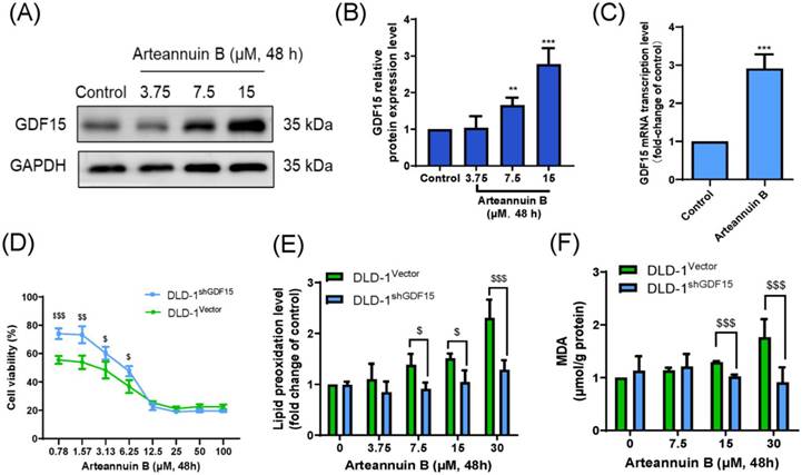
Subsequently, the impact of GDF15 deficiency on the anti-colorectal cancer effect of arteannuin B was studied. We silenced GDF15 in DLD-1 cells using shRNA (Figure S2A-B). Knockdown of GDF15 reduced the sensitivity of DLD-1 cells to arteannuin B, significantly increasing cell viability (Figure 4D). Compared with DLD-1Vector cells, the intracellular lipid peroxidation level of DLD-1shGDF15 cells was significantly reduced (Figure 4E). Similarly, the production rate of MDA also decreased significantly (Figure 4F).
These data suggested that GDF15 was a key factor in arteannuin B-induced ferroptosis in DLD-1 cells. In the absence of GDF15, arteannuin B could not successfully induce intracellular lipid peroxidation, and led to significantly enhanced resistance of DLD-1 cells to arteannuin B.
Arteannuin B significantly inhibited the mevalonate pathway in colorectal cancer
As mentioned earlier, the metabolic flux of MVP is crucial for maintaining the normal function of GPX4 and resisting ferroptosis in cells. The results of KEGG analysis showed that SDEPs were remarkably enriched in MVP (Figure 2D), which is the core metabolic pathway of cholesterol synthesis associated with terpenoid backbone biosynthesis.
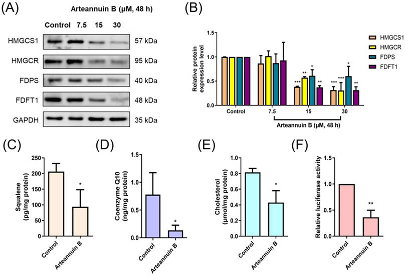
As shown in Figure 5A-B, arteannuin B significantly downregulated the expression of various enzymes in the MVP, including HMGCS1, HMGCR, farnesyl diphosphate synthase (FDPS), and FDFT1. Among them, HMGCS1 and HMGCR are considered as key enzymes of MVP, which directly regulate the flux of MVP [33]. On the other hand, arteannuin B treatment significantly reduced the levels of mevalonate pathway products, including squalene, CoQ10, and cholesterol, in DLD-1 cells (Figure 5C-E). Arteannuin B strongly inhibited the expression of key regulatory enzymes of MVP and led to the reduction of its products. SREBP2 is a classic cholesterol regulatory factor that can directly regulate the gene expression of the mevalonate pathway [34]. The dual luciferase reporter gene experiment showed that arteannuin B can weaken the binding of SREBP2 and HMGCS1(Figure 5F). These results indicate that arteannuin B can significantly inhibit the mevalonate pathway in CRC cells.
Arteannuin B inhibited the mevalonate pathway in colorectal cancer. (A-B) Western blotting images and quantitative analysis showed the effects of arteannuin B on MVP-related protein expression levels in DLD-1 cells. (C-E) Effect of arteannuin B on MVP products in DLD-1 cells, squalene (C), CoQ10 (D) and cholesterol (E). (F) The effect of arteannuin B on the binding of HMGCS1 and SREBP2 (dual luciferase reporter gene assay). Data are presented as mean ± SD, n = 3; *p < 0.05, **p < 0.01, ***p < 0.001 vs. control group.
Arteannuin B induced ferroptosis via GDF15/HMGCS1/GPX4 axis
It has been reported that GDF15 can regulate cholesterol synthesis in esophageal cancer cells through the sterol regulatory element-binding protein cleavage-activating protein (SCAP)/SREBP2 axis [35]. Therefore, we speculated that GDF15 also has a regulatory effect on the cholesterol synthesis pathway in CRC cells. We examined the changes in expression levels of MVP-related enzymes in DLD-1shGDF15 cells. The inhibitory effect of arteannuin B on HMGCS1, instead of HMGCR, was significantly weakened in DLD-1shGDF15 cells (Figure 6A-B). Meanwhile, the inhibitory effect of arteannuin B on cholesterol in DLD-1 cells was reversed when GDF15 was knocked down (Figure 6C). In the absence of GDF15, arteannuin B lost its inhibitory activity against MVP. Knockdown of GDF15 caused significant upregulation of HMGCS1.
Arteannuin B induced ferroptosis via GDF15/HMGCS1/GPX4 axis. (A-B) Western blotting images and quantitative analysis showed the effects of GDF15 knockdown on the expression levels of MVP-related proteins. (C) Effect of GDF15 knockdown on the cholesterol level in DLD-1Vector and DLD-1shGDF15 cells. (D-E) Western blotting images and quantitative analysis showed the effects of arteannuin B on the protein expression level in DLD-1oeHMGCS1 cells. Data are presented as mean ± SD, n = 3; *p < 0.05, **p < 0.01, ***p < 0.001 vs. control group; $p < 0.05, $$p < 0.01 vs. DLD-1Vector cells.
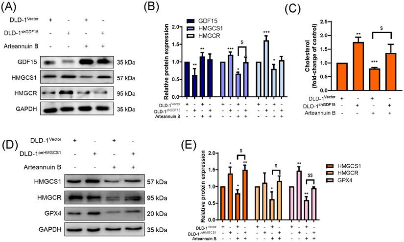
Subsequently, DLD-1 cells with overexpression of HMGCS1 protein were constructed (Figure S2C-D). In DLD-1oeHMGCS1 cells, we observed that the inhibitory effect of arteannuin B on HMGCR and GPX4 was significantly weakened (Figure 6D-E). This result proved that HMGCS1 was a key factor in the regulation of MVP by arteannuin B, and arteannuin B achieved the synthesis inhibition of GPX4 protein by inhibiting HMGCS1.
To further confirm the regulatory effect of GDF15 on MVP, we successfully constructed DLD-1oeGDF15 cells (Figure S2E-F) and examined the effects of GDF15 on MVP and GPX4. The experimental results showed that high-level of GDF15 inhibited the expression of two key enzymes of MVP (HMGCS1 and HMGCR) as well as GPX4 protein (Figure S3A-B). Besides, overexpression of GDF15 formed a synergistic effect with arteannuin B, further inhibiting the expression of the above three proteins.
Our experimental result demonstrated that the arteannuin B-induced ferroptosis and the inhibitory effect of arteannuin B on MVP were all dependent on the presence of GDF15 protein. It supported that arteannuin B induced ferroptosis in CRC cells through the GDF15/HMGCS1/GPX4 signaling axis.
Knockdown of GDF15 weakened the anti-colorectal cancer effect of arteannuin B in vivo
GDF15 has been recognized as a potent suppressor of oncogenic processes, with demonstrated direct interactions with a spectrum of tumor-suppressor genes, including P53, early growth response protein 1 (EGR-1), and peroxisome proliferator-activated receptor gamma (PPARγ). These interactions are posited to contribute to the inhibitory effects of GDF15 on the proliferation of neoplastic cells [36,37]. Using GDF15 and GPX4 expression as variables, we collected the data of patient samples from public databases and conducted Kaplan-Meier survival analyses on the included patient cohort. The GEPIA2 tool was used to construct overall survival curves [38]. The results indicated that high level of GDF15 and low level of GPX4 in tumor tissues typically imply longer median survival (Figure 7A).
Knockdown of GDF15 weakened the anti-colorectal cancer effect of arteannuin B in vivo. (A) Kaplan-Meier analysis of survival statistic in patient data from public databases. The dotted lines represent the 95% confidence intervals (95% CI) for the survival estimates. (B) Schematic diagram of animal experiment. (C) Tumor growth curves (n = 5). (D) Pictures of tumor tissues. (E) Tumor weight (n = 5). (F) Curve of animal weight changes (n = 5). (G-H) Cholesterol and MDA levels in tumor tissues (n = 3). Data are presented as mean ± SD; *p < 0.05, **p < 0.01, ***p < 0.001 vs. model group; #p < 0.05, ##p < 0.01, ###p < 0.001 vs. DLD-1WT cells; ns, no significant.
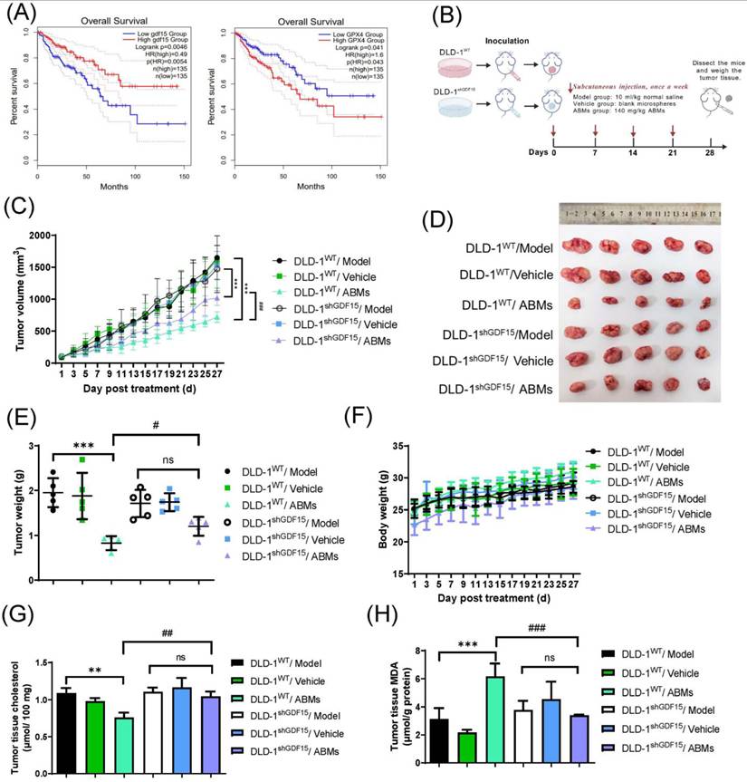
Next, we investigated the impact of GDF15 knockdown on the in vivo anti-colorectal cancer efficacy of arteannuin B (Figure 7B). ABMs were prepared as our previous report [39]. The results of xenograft tumor experiment indicated that ABMs significantly inhibited the growth of DLD-1WT and DLD-1shGDF15 cells in vivo (Figure 7C-D). DLD-1WT/Model group and DLD-1shGDF15/Model group showed similar growth curves indicating that knockdown of GDF15 did not affect the growth of DLD-1 xenograft tumors. During the administration cycle, the loss of GDF15 protein significantly reduced the sensitivity of DLD-1 cells to ABMs, weakening the inhibition of xenograft tumor growth by ABMs (Figure 7C, DLD-1 shGDF15/ABMs group vs DLD-1WT/ABMs group). After the tumor tissue was dissected and the tumor inhibition rate was calculated, the results showed that the inhibitory rates of ABMs on DLD-1WT and DLD-1shGDF15 xenograft tumors were 57.64% (DLD-1WT/ABMs group) and 29.71% (DLD-1shGDF15/ABMs group), respectively (Figure 7E). The in vivo anti-colorectal cancer effect of arteannuin B was greatly inhibited in the absence of GDF15.
During the administration period, there were no significant changes between ABMs groups and model groups in the body weight (Figure 7F) and the organ indices of mice (Table 2).
Organ indices of the nude mice xenografted with DLD-1WT or DLD-1shGDF15 cells.
| Groups | Organ Indices (mg/g, x̄ ± SD, n = 5) | |||
|---|---|---|---|---|
| Heart | Liver | Spleen | Kidney | |
| DLD-1WT/Model | 7.52 ± 1.07 | 53.03 ± 7.45 | 4.93 ± 0.63 | 16.18 ± 0.6 |
| DLD-1WT/Vehicle | 7.53 ± 1.45 | 52.01 ± 6.58 | 4.82 ± 0.73 | 16.01 ± 0.73 |
| DLD-1WT/ABMs | 7.34 ± 0.53 | 53.23 ± 9.10 | 6.07 ± 1.34 | 16.20 ± 1.95 |
| DLD-1shGDF15/ Model | 7.90 ± 0.98 | 56.80 ± 5.02 | 4.76 ± 0.8 | 17.56 ± 1.20 |
| DLD-1shGDF15/Vehicle | 8.10 ± 1.38 | 54.76 ± 4.58 | 4.58 ± 0.57 | 15.78 ± 2.45 |
| DLD-1shGDF15/ABMs | 7.76 ± 1.48 | 56.13 ± 4.52 | 5.81 ± 0.93 | 17.64 ± 1.09 |
IHC analysis was used to examine the protein expression of GPX4 in tumor tissues. As shown in Figure S4, arteannuin B significantly inhibited the protein expression of GPX4 in DLD-1WT xenograft tumor tissues. However, the protein expression of GPX4 was restored in DLD-1shGDF15 xenograft tumor tissues.
Similarly, we also observed that the level of cholesterol, the end-product of MVP, was significantly inhibited by arteannuin B in DLD-1WT xenograft tumor tissues. This inhibitory effect was eliminated in DLD-1shGDF15 xenograft tumor tissues (Figure 7G). On the other hand, arteannuin B treatment induced an increase in the content of lipid peroxidation product MDA in tumor tissues, and this phenomenon was alleviated by knockdown of GDF15 (Figure 7H).
The results above demonstrated that GDF15 is a key protein in arteannuin B-induced ferroptosis in CRC cells in vitro and in vivo. The anti-colorectal cancer effect of arteannuin B was greatly weakened after GDF15 was knocked down.
Discussion
At present, the standard treatments for colorectal cancer include surgery, radiotherapy, and chemotherapy [40]. Nevertheless, the issue of drug resistance has underscored an urgent call for the development of novel therapeutic agents. In this study, we evaluated the anti-tumor activity of arteannuin B, a sesquiterpene lactone in Artemisia annua, both in vitro and in vivo. For the first time, we systematically demonstrated that arteannuin B induces ferroptosis in CRC cells.
Previous studies have established artemisinin compounds as ferroptosis inducers, which primarily regulate Fe²⁺ metabolism and oxidative stress [41], while alternative mechanisms include endoplasmic reticulum stress [42], P53 signaling [43], regulation of SREBP2 nuclear localization [44]. Our findings indicated that arteannuin B was more likely to disrupt intracellular lipid metabolism (Figure 3B-D), rather than directly perturb Fe2+ metabolism (Figure 3E). We proposed a novel mechanism by which arteannuin B induces ferroptosis in CRC cells through the GDF15/HMGCS1/GPX4 axis.
GDF15 was identified as the most significantly upregulated protein after treatment with arteannuin B (Figure 2C). When GDF15 was knocked down, arteannuin B-induced ferroptosis was weakened both in vitro and in vivo (Figure 4D-F and Figure 7C-E), and tumor tissue cholesterol no longer decreased (Figure 7G), indicating that GDF15 is a necessary upstream switch for inhibiting the mevalonate pathway and subsequent ferroptosis.
Recent studies have reported conflicting roles of GDF15 in ferroptosis. In microsatellite instability-high colorectal cancer (MSI-H CRC), GDF15 suppression enhances ferroptosis sensitivity [45], whereas in ccRCC, GDF15 overexpression promotes ferroptosis [16]. Another study showed that GDF15 overexpression alleviates myocardial ischemia-reperfusion injury by inhibiting ferroptosis [46]. In contrast, our findings demonstrate that arteannuin B upregulates GDF15 and this induction is essential for ferroptosis in CRC cells. These discrepancies strongly suggest that GDF15's role in ferroptosis is highly context-dependent, varying across tumor types, molecular subtypes, and disease settings.
Mechanistically, our study reveals that GDF15 suppresses the mevalonate pathway by downregulating HMGCS1, which subsequently leads to the reduction of GPX4. In contrast, Huang et al. [45] found that GDF15 knockdown reduces solute carrier family 7 member 11 (SLC7A11) expression, thereby promoting ferroptosis. This suggests that GDF15 may regulate ferroptosis through multiple axes, depending on the cellular context. The opposing roles of GDF15 may also reflect treatment context: our study examines GDF15 induction by arteannuin B, whereas Huang et al. investigated baseline GDF15 suppression. Thus, GDF15 may function as a sensor of metabolic stress, exerting pro- or anti-ferroptosis effects depending on the triggering stimulus.
To our knowledge, this is the first study to show that pharmacological induction of GDF15 by arteannuin B can sensitize CRC cells to ferroptosis, revealing a therapeutic avenue distinct from GDF15's endogenous roles in MSI-H CRC or ccRCC. However, a key limitation of our work is that all mechanistic experiments were conducted in a single CRC cell line (DLD-1). Future studies should validate these results in additional CRC models, including patient-derived organoids or in vivo systems representing diverse molecular subtypes, to better define the scope and translational relevance of the GDF15-ferroptosis axis.
In addition, our study demonstrated that arteannuin B regulates ferroptosis via the HMGCS1-mevalonate pathway. TMT proteomics revealed that 48 h treatment with 15 μM arteannuin B markedly reduced the mevalonate-pathway gatekeeper HMGCS1 (Figure 2C), and consequently, subsequent cellular assays confirmed the decrease in its downstream products CoQ10 and cholesterol (Figure 5D-E). In DLD-1oeHMGCS1 cells, the downregulation of GPX4 expression induced by arteannuin B was reverted to control levels (Figure 6D-E). In DLD-1shGDF15 cells, the elevated lipid peroxidation and MDA levels caused by arteannuin B were substantially reduced (Figure 4E-F), and both HMGCS1 and cholesterol levels were restored (Figure 6A-C). These data suggest that arteannuin B blocks the mevalonate pathway through HMGCS1 rather than the classical target HMGCR, thereby inhibiting GPX4 and triggering ferroptosis. This finding points out that the mevalonate pathway is involved in ferroptosis not only through the HMGCR/GPX4 axis (like statins [47]), offering a new molecular target for mevalonate pathway-directed anticancer strategies.
Although our study provides compelling evidence for the anti-cancer mechanism of arteannuin B in colorectal cancer, certain limitations should be acknowledged. Arteannuin B as an ferroptosis inducer is a promising but still in early research. Although our in vitro screening encompassed a panel of nine cell lines, the detailed mechanistic dissection was primarily focused on the DLD-1 model, which exhibited the highest sensitivity to the compound. Future investigations involving a broader spectrum of molecular subtypes would further consolidate the universality of the GDF15-mediated pathway. Furthermore, regarding the preclinical models, although the subcutaneous xenograft data strongly support the in vivo efficacy, the incorporation of more complex models, such as patient-derived xenografts (PDX) or orthotopic models, would provide a more comprehensive evaluation of its therapeutic potential. Finally, while our preliminary toxicity assessments, demonstrating stable body weight and organ indices, suggest a favorable safety profile, it would be more compelling to include a thorough toxicological analysis, including hematology and histopathology, in future studies to fully pave the way for clinical translation.
Conclusion
In conclusion, our findings confirm that arteannuin B triggers ferroptosis-like cell death in CRC cells and suppresses xenograft growth. This anti-tumor effect is in association with inhibition of the mevalonate pathway, leading to the disruption of redox homeostasis. Notably, we elucidate that GDF15 contributes to arteannuin B-mediated suppression of HMGCS1 and GPX4, establishing GDF15 as an essential sensitizer for ferroptosis induction. These results provide a mechanistic rationale for targeting the GDF15-mevalonate pathway axis in CRC therapy.
Supplementary Material
Supplementary figures and tables.

Abbreviations
3-MA: 3-methyladenine; 5-FU: 5-fluorouracil; 7-DHC: 7-dehydrocholesterol; BCA: bicinchoninic acid; ccRCC: clear cell renal cell carcinoma; CoQ10: coenzyme Q10; DCFH-DA: 2,7-dichlorodihydrofluorescein diacetate; DMSO: dimethyl sulfoxide; EGR-1: early growth response protein 1; FDFT1: farnesyl diphosphate farnesyltransferase 1; FDPS: farnesyl diphosphate synthase; Fer-1: ferrostatin-1; GDF15: growth differentiation factor 15; GPX4: glutathione peroxidase 4; HMGCR: 3-hydroxy-3-methylglutaryl-CoA reductase; HMGCS1: 3-hydroxy-3-methylglutaryl-CoA synthase 1; IHC: immunohistochemistry; KEGG: Kyoto Encyclopedia of Genes and Genomes; MDA: malondialdehyde; MVP: mevalonate pathway; Nec-1: necrostatin-1; PPARγ: peroxisome proliferator-activated receptor gamma; ROS: reactive oxygen species; SCAP: sterol regulatory element-binding protein cleavage-activating protein; SDEPs: significantly differentially expressed proteins; SLC7A11: solute carrier family 7 member 11; SREBP2: sterol regulatory element-binding protein 2; TMT: tandem mass tag; Z-VAD-FMK: benzyloxycarbonyl-Val-Ala-Asp(OMe)-fluoromethylketone.
Acknowledgements
The Figure 7B in this article were created with BioGDP.com [48].
Funding
This work was funded by National Natural Science Foundation of China [Nos. 82174019, 82003774 and 81673646].
Data availability
The data supporting the findings of this study are available from the corresponding author upon reasonable request.
Ethical approval
The animal experiments described in this article were approved by the Experimental Animal Ethics Committee of Jinan University and conducted in accordance with the principles of animal protection, animal welfare, and experimental ethics. The approval number for the in vivo anti-colorectal cancer experiment using arteannuin B is IACUC-2019191014-08. The approval number for the in vivo experiment aimed at identifying GDF15 as a key target protein is IACUC-20230812-06.
Informed consent
All participants gave written informed consent before undergoing any study-related procedures.
Author contributions
X.Z.O. and L.Y.S. conceived the study and designed the methodology. X.Z.O., J.H.X. and Y.T.J. performed the experiments, collected and curated the data, and carried out validation. Y.Q.W. provided the necessary resources. R.M.Y., J.H.Z., W.J.H. and L.Y.S. jointly supervised the project, administered it, and secured funding. X.Z.O. wrote the original draft. X.Z.O., J.H.X., Y.T.J. and L.Y.S. reviewed and edited the manuscript. All authors contributed to the article and approved the submitted version.
Competing Interests
The authors have declared that no competing interest exists.
References
1. Siegel RL, Wagle NS, Cercek A. et al. Colorectal cancer statistics, 2023. CA Cancer J Clin. 2023;73:233-254
2. Morgan E, Arnold M, Gini A. et al. Global burden of colorectal cancer in 2020 and 2040: incidence and mortality estimates from GLOBOCAN. Gut. 2023;72:338-344
3. Fong W, To KKW. Drug repurposing to overcome resistance to various therapies for colorectal cancer. Cell Mol Life Sci. 2019;76:3383-3406
4. Dixon SJ, Lemberg KM, Lamprecht MR. et al. Ferroptosis: an iron-dependent form of nonapoptotic cell death. Cell. 2012;149:1060-1072
5. Yang WS, SriRamaratnam R, Welsch ME. et al. Regulation of ferroptotic cancer cell death by GPX4. Cell. 2014;156:317-331
6. Sun Q, Liu D, Cui W. et al. Cholesterol mediated ferroptosis suppression reveals essential roles of Coenzyme Q and squalene. Commun Biol. 2023 6
7. Bersuker K, Hendricks JM, Li Z. et al. The CoQ oxidoreductase FSP1 acts parallel to GPX4 to inhibit ferroptosis. Nature. 2019;575:688-692
8. Friedmann Angeli JP, Conrad M. Selenium and GPX4, a vital symbiosis. Free Radical Biol. Med. 2018;127:153-159
9. Yang WS, Stockwell BR. Ferroptosis: death by lipid peroxidation. Trends Cell Biol. 2016;26:165-176
10. Kitsugi K, Noritake H, Matsumoto M. et al. Simvastatin inhibits hepatic stellate cells activation by regulating the ferroptosis signaling pathway. Biochim Biophys Acta - Mol Basis Dis. 2023;1869:166750
11. Shu X, Wu J, Zhang T. et al. Statin-induced geranylgeranyl pyrophosphate depletion promotes ferroptosis-related senescence in adipose tissue. Nutrients. 2022;14:4365
12. Li Y, Ran Q, Duan Q. et al. 7-Dehydrocholesterol dictates ferroptosis sensitivity. Nature. 2024;626:411-418
13. Dong X, Xu D. Research progress on the role and mechanism of GDF15 in body weight regulation. Obesity Facts. 2024;17:1-11
14. Dixon SJ, Patel DN, Welsch M. et al. Pharmacological inhibition of cystine-glutamate exchange induces endoplasmic reticulum stress and ferroptosis. elife. 2014 3
15. Shao Y, Jia H, Huang L. et al. An original ferroptosis-related gene signature effectively predicts the prognosis and clinical status for colorectal cancer patients. Front Oncol. 2021 11
16. Yang D, He Z, Lu J. et al. Downregulation of GDF15 suppresses ferroptosis and predicts unfavorable prognosis in clear cell renal cell carcinoma. Cell Div. 2023 18
17. Efferth T, Kaina B. Toxicity of the antimalarial artemisinin and its derivatives. Crit Rev Toxicol. 2010;40:405-421
18. Feng X, Cao S, Qiu F. et al. Traditional application and modern pharmacological research of Artemisia annua L. Pharmacol Ther. 2020;216:107650
19. Emadi F, Yassa N, Hadjiakhoondi A. et al. Sedative effects of Iranian Artemisia annua in mice: Possible benzodiazepine receptors involvement. Pharm Biol. 2011;49:784-788
20. Lee SK, Kim H, Park J. et al. Artemisia annua extract prevents ovariectomy-induced bone loss by blocking receptor activator of nuclear factor kappa-B ligand-induced differentiation of osteoclasts. Sci Rep. 2017 7
21. Chen H, Hu Q, Wen T. et al. Arteannuin B, a sesquiterpene lactone from Artemisia annua, attenuates inflammatory response by inhibiting the ubiquitin-conjugating enzyme UBE2D3-mediated NF-kappa B activation. Phytomedicine. 2024;124:155263
22. Sawhney G, Rasool JU, Saroch D. et al. Arteannuin-B and (3-Chlorophenyl)-2-Spiroisoxazoline Derivative Exhibit Anti-Inflammatory Effects in LPS-Activated RAW 264.7 Macrophages and BALB/c Mice-Induced Proinflammatory Responses via Downregulation of NF-κB/P38 MAPK Signaling. Molecules. 2022;27:8068
23. Hu Y, Liu M, Qin H. et al. Artemether, artesunate, arteannuin B, echinatin, licochalcone B and andrographolide effectively inhibit SARS-CoV-2 and related viruses in vitro. Front Cell Infect Microbiol. 2021 11
24. Varela K, Arman HD, Berger MS. et al. Inhibition of cysteine proteases via thiol-michael addition explains the anti-SARS-CoV-2 and bioactive properties of arteannuin B. J Nat Prod. 2023;86:1654-1666
25. Efferth T, Herrmann F, Tahrani A. et al. Cytotoxic activity of secondary metabolites derived from Artemisia annua L. towards cancer cells in comparison to its designated active constituent artemisinin. Phytomedicine. 2011;18:959-969
26. Huang W, Wang Y, He T. et al. Arteannuin B enhances the effectiveness of cisplatin in non-small cell lung cancer by regulating connexin 43 and MAPK pathway. Am J Chin Med. 2022;50:1963-1992
27. Hu Y, Guo N, Yang T. et al. The potential mechanisms by which artemisinin and its derivatives induce ferroptosis in the treatment of cancer. Oxid. Med. Cell. Longevity. 2022;2022:1-12
28. Nishizawa H, Matsumoto M, Shindo T. et al. Ferroptosis is controlled by the coordinated transcriptional regulation of glutathione and labile iron metabolism by the transcription factor BACH1. J Biol Chem. 2020;295:69-82
29. Yang WS, Kim KJ, Gaschler MM. et al. Peroxidation of polyunsaturated fatty acids by lipoxygenases drives ferroptosis. Proc Natl Acad Sci U S A. 2016 113
30. Luo L, Deng L, Chen Y. et al. Identification of lipocalin 2 as a ferroptosis-related key gene associated with hypoxic-ischemic brain damage via STAT3/NF-ΚB signaling pathway. Antioxidants (Basel). 2023;12:186
31. Lee SH, Krisanapun C, Baek SJ. NSAID-activated gene-1 as a molecular target for capsaicin-induced apoptosis through a novel molecular mechanism involving GSK3, C/EBP and ATF3. Carcinogenesis. 2010;31:719-728
32. Shin SY, Kim JH, Lee JH. et al. 2′-Hydroxyflavanone induces apoptosis through Egr-1 involving expression of Bax, p21, and NAG-1 in colon cancer cells. Mol Nutr Food Res. 2012;56:761-774
33. Wang I, Huang T, Chen J. et al. Mevalonate pathway enzyme HMGCS1 contributes to gastric cancer progression. Cancers. 2020;12:1088
34. Wong JVS, Fatehi Hassanabad A. Targeting the Mevalonate Pathway for Treating Esophageal Cancer. J Gastrointest Canc. 2021;52:819-821
35. Dong G, Huang X, Jiang S. et al. SCAP Mediated GDF15-Induced Invasion and EMT of Esophageal Cancer. Front Oncol. 2020 10
36. Lee J, Kim I, Yoo E. et al. Competitive inhibition by NAG-1/GDF-15 NLS peptide enhances its anti-cancer activity. Biochem Biophys Res Commun. 2019;519:29-34
37. Wang X, Baek SJ, Eling TE. The diverse roles of nonsteroidal anti-inflammatory drug activated gene (NAG-1/GDF15) in cancer. Biochem Pharmacol. 2013;85:597-606
38. Tang Z, Kang B, Li C. et al. GEPIA2: an enhanced web server for large-scale expression profiling and interactive analysis. Nucleic Acids Res. 2019;47:W556-W560
39. Wang Y, Huang W, Wang N. et al. Development of arteannuin B sustained-release microspheres for anti-tumor therapy by integrated experimental and molecular modeling approaches. Pharmaceutics. 2021;13:1236
40. Dekker E, Tanis PJ, Vleugels JLA. et al. Colorectal cancer. The Lancet. 2019;394:1467-1480
41. Grignano E, Cantero-Aguilar L, Tuerdi Z. et al. Dihydroartemisinin-induced ferroptosis in acute myeloid leukemia: links to iron metabolism and metallothionein. Cell Death Dis. 2023;9:97
42. Wang N, Zeng G, Yin J. et al. Artesunate activates the ATF4-CHOP-CHAC1 pathway and affects ferroptosis in Burkitt's Lymphoma. Biochem Biophys Res Commun. 2019;519:533-539
43. Wang L, Zhang Z, Li M. et al. P53-dependent induction of ferroptosis is required for artemether to alleviate carbon tetrachloride-induced liver fibrosis and hepatic stellate cell activation. IUBMB Life. 2019;71:45-56
44. Liang L, Liu Y, Wu X. et al. Artesunate induces ferroptosis by inhibiting the nuclear localization of SREBP2 in myeloma cells. Int J Med Sci. 2023;20:1535-1550
45. Huangfu ZM, Xiao M, Xie H. et al. Suppressing GDF15 enhances the chemotherapeutic effect of 5-FU on MSI-H CRC by regulating the ferroptosis pathway SLC7A11/GSH/GPX4. J Cancer Res Clin Oncol. 2025;151:6
46. Gao Q, Li C, Zhong P. et al. GDF15 restrains myocardial ischemia-reperfusion injury through inhibiting GPX4 mediated ferroptosis. Aging (Albany NY). 2024;16:617-626
47. Yao X, Xie R, Cao Y. et al. Simvastatin induced ferroptosis for triple-negative breast cancer therapy. J Nanobiotechnol. 2021;19:311
48. Jiang S, Li H, Zhang L. et al. Generic Diagramming Platform (GDP): a comprehensive database of high-quality biomedical graphics. Nucleic Acids Res. 2025;53:D1670-D1676
Author biography
Dr. Xiaozheng Ou obtained her PhD in Microbiology and Biochemistry Pharmacy from Jinan University (Guangzhou, China) and collaborated with Professor Liyan Song and Professor Weijuan Huang to develop new cancer treatment drugs. She is currently a researcher at the Second Affiliated Hospital of South China University of Technology.
Dr. Liyan Song is a professor of cancer pharmacology at the School of Pharmacy, Jinan University. She has co-authored 17 SCI-indexed publications and holds four Chinese patents. Prof. Song has led or participated in more than 10 national projects funded by the NSFC and National Key R&D Program. Current research in her group focuses on: (1) tumor immunopharmacology and mechanisms of traditional Chinese medicine; (2) screening and validation of active anti-cancer compounds from marine and herbal medicines; and (3) translational development of innovative small-molecule or biologic drugs.
Corresponding authors: Liyan Song (E-mail: tslyedu.cn) and Weijuan Huang (wjhuangedu.cn), Biotechnological Institute of Chinese Materia Medica, Jinan University, 855 Xingye Avenue East, Guangzhou 511443, China. Fax: (+86) 20-8522-8205.