Impact Factor ISSN: 1837-9664




J Cancer 2026; 17(8):1435-1444. doi:10.7150/jca.136981 This issue Cite
Research Paper
Selective sensitivity of betel nut-related oral squamous cell carcinoma to evodiamine through cell cycle arrest, growth inhibition and apoptosis induction
Yin-Che Lu1, Kai-Liang Tang2, Chih-Hsien Liao3, Yi-Zhen Li4, Chin-Ho Kuo1, Pin-Tzu Chen1, Shu-Hsin Chen3, Ying-Ray Lee5,6,7,8,9,10 
1. Division of Hematology-Oncology, Ditmanson Medical Foundation Chia-Yi Christian Hospital, Chiayi, Taiwan.
2. Taichung District Agricultural Research and Extension station, Ministry of Agriculture, Changhua County, Taiwan.
3. Department of Medical Research, Ditmanson Medical Foundation Chiayi Christian Hospital, Chiayi, Taiwan.
4. Bone and Joint Research Center, Chang Gung Memorial Hospital, Taoyuan, Taiwan.
5. School of Medicine, College of Medicine, Kaohsiung Medical University, Kaohsiung, Taiwan.
6. Department of Microbiology and Immunology, College of Medicine, Kaohsiung Medical University, Kaohsiung, Taiwan.
7. Master of Science Program in Tropical Medicine, College of Medicine, Kaohsiung Medical University, Kaohsiung, Taiwan.
8. Faculty of Post-Baccalaureate Medicine, College of Medicine, Kaohsiung Medical University, Kaohsiung, Taiwan.
9. Center for Tropical Medicine and Infectious Disease, Kaohsiung Medical University, Kaohsiung, Taiwan.
10. Department of Medical Research, Kaohsiung Medical University Hospital, Kaohsiung, Taiwan.
Received 2026-4-29; Accepted 2026-7-23; Published 2026-7-30
Abstract
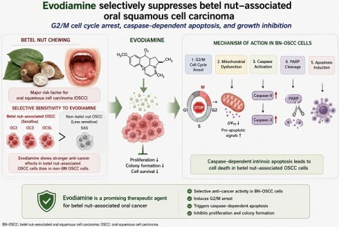
Head and neck cancer, particularly oral squamous cell carcinoma (OSCC), remains a major global health burden, with especially high incidence and mortality in Taiwan due to the widespread habit of betel quid chewing. Betel nut mediated OSCC (BN-OSCC) is characterized by aggressive clinical behavior, including increased risks of recurrence, metastasis, and poor survival outcomes, and exhibits limited responsiveness to conventional chemoradiotherapy. Therefore, the development of novel therapeutic agents and radiosensitizers is urgently needed.
Evodiamine (EVO), a bioactive quinolone alkaloid isolated from Evodia rutaecarpa, has demonstrated broad anti-tumor activities across multiple cancer types, including inhibition of proliferation, suppression of metastasis, and induction of apoptosis. Although previous studies have reported the anti-OSCC effects of EVO through modulation of pathways such as Akt, EPRS1, and RAGE, its specific impact on BN-OSCC remains unclear.
In this study, we investigated the anti-cancer effects of EVO in BN-OSCC and non-BN-OSCC cell lines. Our results demonstrate that EVO significantly inhibits cell viability and proliferation in a dose-dependent manner in BN-OSCC rather than non-BN-OSCC cells. Importantly, EVO induces apoptosis through activation of the caspase-dependent pathway, as evidenced by increased cleavage of caspase-3, caspase-9, and PARP. Furthermore, EVO treatment leads to mitochondrial dysfunction and enhances pro-apoptotic signaling, suggesting involvement of the intrinsic apoptotic pathway.
Collectively, these findings indicate that EVO exerts potent anti-tumor effects in BN-OSCC cells by triggering caspase-dependent apoptosis. This study provides novel insights into the therapeutic potential of EVO and supports its development as a promising candidate for the treatment of betel nut mediated oral cancer.
Keywords: evodiamine, anti-cancer, OSCC, apoptosis
Introduction
Head and neck cancers, including malignancies of the oral cavity, such as the buccal mucosa, gingiva, and tongue, represent a major global health burden. These cancers rank the sixth common malignancy worldwide, with approximately 650,000 new cases and 200,000 deaths annually. In the USA, nearly 54,000 new cases are diagnosed each year, of which more than 99% are histologically classified as squamous cell carcinomas [1]. Notably, in Taiwan, head and neck cancers are the fourth leading cause of cancer-related mortality and the sixth most prevalent cancer overall [2], underscoring its significant regional impact.
Although tobacco smoking and alcohol consumption are considered well-established risk factors for oral carcinogenesis [3], epidemiological evidence has shown that betel quid chewing is a predominant and independent etiological factor in Taiwan and across Southeast Asia [4-6]. Individuals who chew betel nut exhibit a markedly increased risk of local recurrence, distant metastasis, and developing secondary primary tumors, as well as significantly poorer disease-specific and overall survival compared with non-chewers [4]. In Taiwan, the majority of patients with head and neck cancers are male betel quid users, and a substantial proportion presents with an advanced locoregional disease (stages III-IV). The current standard treatment for these patients typically involves multimodal strategies, including chemotherapy combined with radiotherapy (RT). However, despite aggressive treatment, the five-year survival rate remains below 50% because of intrinsic or acquired therapeutic resistance [7, 8]. Therefore, identifying effective therapeutic agents and radiosensitizers for betel nut-associated oral squamous cell carcinoma (BN-OSCC) is urgently necessary.
Natural compounds derived from traditional Chinese medicinal herbs have attracted increasing attention as potential anticancer agents and adjuvants that can enhance the efficacy of conventional therapies [9]. Evodiamine (EVO), a quinolone alkaloid isolated from Evodia rutaecarpa Bentham, has been extensively studied for its broad spectrum of pharmacological activities, including anti-inflammatory, antimicrobial, antiobesity, neuroprotective, and antiviral effects [10-12]. Accumulating evidence has shown that EVO possesses potent antitumor properties across a wide range of human malignancies, including cancers of the esophagus, lung, breast, liver, colon, prostate, bladder, pancreas, stomach, ovary, and thyroid, as well as melanoma, glioblastoma, and T-cell lymphoma [9, 13-17]. Mechanistically, EVO has been shown to inhibit tumor cell proliferation, induce cell cycle arrest, suppress metastasis, and trigger apoptosis by modulating multiple signaling pathways [13]. Furthermore, EVO has been reported to target cancer stem cells, inhibit epithelial-mesenchymal transition (EMT), and overcome drug resistance, thereby highlighting its therapeutic potential [13].
In the context of OSCC, several studies have reported that EVO exerts antitumor effects by inhibiting cell proliferation and inducing apoptosis [18-21]. These effects are partly mediated through the modulation of key signaling pathways, including the Akt pathway, glutamyl-prolyl-tRNA synthetase 1 (EPRS1), and the receptor for advanced glycation-end products (RAGE). In addition, EVO has been shown to suppress invasion and angiogenesis in OSCC cells via RAGE-dependent mechanisms [18, 20, 21]. Despite these promising findings, the specific effects and underlying mechanism of EVO in BN-OSCC remain unknown.
Therefore, the present study aimed to investigate the antitumor activity of EVO in BN-OSCC cells, with a particular focus on its potential application as a therapeutic agent and radiosensitizer in this high-risk and clinically challenging subtype of oral cancer.
Materials and Methods
Cell lines and cell culture
Four human OSCC cell lines, including SAS, OC2, OC3, and OCSL, were employed in this study. The SAS cell line was obtained from the Japanese Collection of Research Bioresources Cell Bank and cultured in Dulbecco's modified Eagle medium (Gibco, Grand Island, NY, USA) containing 10% fetal bovine serum (FBS) and 1% penicillin-streptomycin. The OC2, OC3, and OCSL cell lines, which were originally established from Taiwanese male patients with histories of alcohol consumption, cigarette smoking, and betel quid chewing [6], were maintained in an RPMI-1640 medium supplemented with 10% FBS and 1% penicillin-streptomycin. All cell lines were incubated at 37 °C in a humidified atmosphere containing 5% CO₂.
Cell viability analysis
Cell viability was assessed using the CCK-8 assay (Enzo Life Sciences, Farmingdale, NY, USA). In brief, 5 × 10³ cells were seeded into a 96-well plate and treated with different concentrations of EVO (0.5-5 μM; Sigma-Aldrich; St. Louis, MO, USA) or a vehicle control (0.01% DMSO) for the indicated durations (24-72 h). Following treatment, a CCK-8 reagent was added and incubated in accordance with the manufacturer's protocol. Absorbance was measured using a microplate reader, and cell viability was expressed relative to the vehicle-treated control group. Each experiment was conducted in triplicate and independently repeated at least three times.
Colony formation assay
For colony formation analysis, cells were seeded into six-well plates at 1 × 10³ cells per well and maintained under standard culture conditions at 37 °C. Following overnight attachment, the cells were exposed to either a vehicle control medium (0.01% DMSO) or a medium containing EVO for 24 h. After treatment, the culture medium was replaced, and the cells were allowed to grow for an additional 10 days. Colonies were subsequently fixed and stained with a 10% crystal violet solution (Sigma-Aldrich). Colony morphology was documented, and colony number and size were quantified.
Cell cycle analysis
To determine the effects of EVO on cell cycle progression, cells were treated with a vehicle control or increasing concentrations of EVO for the indicated time periods (24-72 h). After treatment, the cells were harvested and preserved in cold 70% ethanol overnight. The fixed samples were rinsed two times with PBS and stained with a PI solution (Sigma-Aldrich) at room temperature for 30 min under light-protected conditions. Cellular DNA content was quantified by flow cytometry using a FACScan instrument (Becton Dickinson, San Diego, CA, USA), and the proportion of cells in different cell-cycle phases were calculated using ModFit LT 3.3 software. Each experiment was repeated independently on three separate occasions.
Cellular apoptosis analysis
Apoptosis was quantified by Annexin V-FITC/PI double staining and flow cytometry [14, 22, 23]. Cells were treated with EVO or a vehicle control for the indicated concentrations and incubation periods. Afterward, cells were harvested, washed two times with PBS, and resuspended in a binding buffer containing Annexin V-FITC and PI. After incubation for 15 min in the dark at room temperature, fluorescence signals were measured using a FACScan flow cytometer (Becton Dickinson). For each sample, 1 × 10⁵ cells were analyzed, and the percentage of apoptotic cells was calculated. Three independent experiments were conducted to ensure reproducibility.
Western blotting
The involvement of apoptotic signaling pathways in response to EVO treatment was evaluated using Western blot analysis. The cells exposed to a vehicle control or EVO were harvested, and the total cellular proteins were extracted. Protein samples were resolved by SDS-PAGE and electrotransferred onto PVDF membranes. The membranes were blocked and sequentially incubated with primary and secondary antibodies. The expression levels of apoptosis-associated proteins, including caspase-3 (#9662, Cell Signaling, Danvers, MA, USA), caspase-8 (#9746, Cell Signaling), and caspase-9 (#9502, Cell Signaling), were analyzed, and GAPDH (GTX100118, GeneTex, Hsinchu City, Taiwan) was used as a loading control. Immunoreactive signals were visualized and quantified in accordance with standard Western blotting procedures.
To clarify the role of caspases in EVO-triggered apoptosis, cells were treated with the pan-caspase inhibitor Z-VAD-FMK (BioVision, Mountain View, CA, USA) before EVO administration. Subsequently, the extent of apoptosis was evaluated by Annexin V-FITC and PI co-staining followed by flow cytometric detection. Comparisons between the inhibitor-treated and untreated groups were used to determine the contribution of caspase activation to EVO-induced cell death.
Statistical analysis
All quantitative data were obtained from at least three independent experiments and are presented as the mean ± SD. Statistical analyses were conducted using GraphPad Prism 8 (GraphPad Software, USA). The differences among experimental groups were assessed by one-way ANOVA with Fisher's LSD post hoc analysis. Statistical significance was defined as a two-tailed P value of < 0.05. The levels of significance are indicated as P < 0.05, P < 0.01, and P < 0.001 throughout the study.
Results
Evodiamine inhibits the proliferation of human non-BN-OSCC and BN-OSCC cells
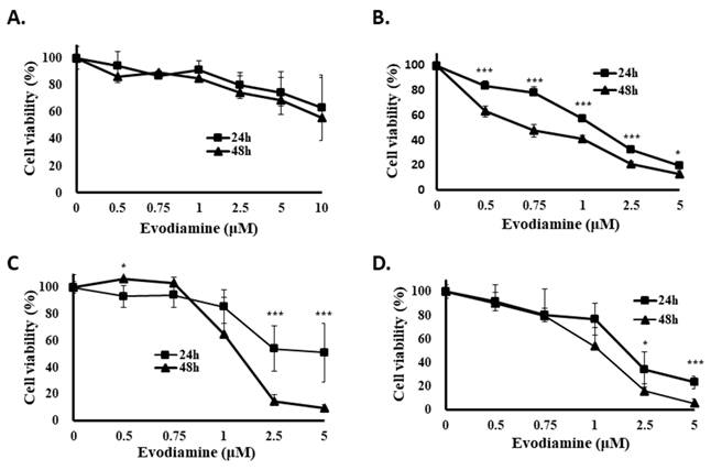
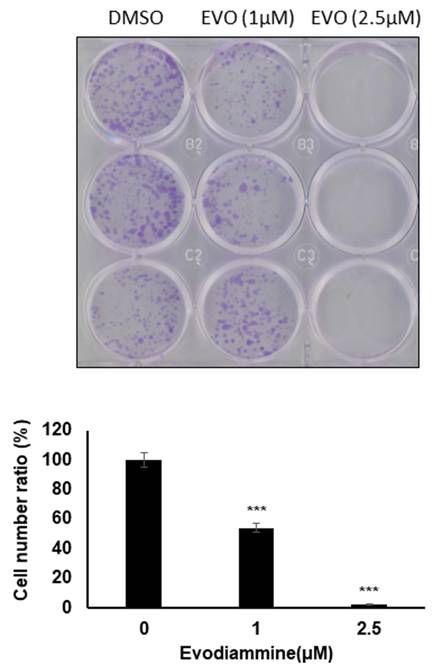
Human non-BN-OSCC cell lines, SAS cells, and BN-OSCC cell lines, including OC2, OC3, and OCSL cells, were used to examine the potential effect of EVO. The growth inhibition of OC2, OC3, and OCSL cells was observed in a dosage- and time-dependent manner after EVO treatment (Fig. 1). However, the effect of EVO on growth inhibition resistance in SAS cells was greater than that in BN-OSCC cell lines (Fig. 1). The IC₅₀ value of EVO in SAS cells was not detectable. By contrast, EVO exhibited significant cytotoxicity in BN-OSCC cell lines. In particular, the IC₅₀ values in OC2 cells were 1.45 ± 0.02 μM and 0.71 ± 0.03 μM after 24 and 48 h of treatment, respectively. In OC3 cells, the IC₅₀ values were 2.51 ± 0.16 μM and 1.44 ± 0.10 μM after 24 and 48 h of treatment, respectively. On the contrary, the IC₅₀ values in OCSL cells were 1.94 ± 0.09 μM and 1.15 ± 0.08 μM after 24 and 48 h of treatment, respectively. In addition, the antiproliferative effect of EVO was evaluated in OC2 cells using a colony formation assay, which confirmed that EVO effectively inhibits the proliferation of BN-OSCC cells (Fig. 2). These results indicate that EVO selectively suppresses cell proliferation in human BN-OSCC cells but not in OCSS cells, highlighting its potential application as a therapeutic agent for BN-OSCC.
Evodiamine suppresses cell proliferation in human BN-OSCC cells rather than OSCC cells. (A) Human OSCC cell line (SAS), and BN-OSCC cell lines (B: OC2, C: OC3, and D: OCSL) were incubated with DMSO or EVO, and the cell viability was determined with CCK-8 assay. Three independent experiments were conducted.
Evodiamine suppresses BN-OSCC cells proliferation. OC2 cells were incubated with evodiamine or DMSO for 24 h, and the colony formation was determined with crystal violet staining after 10days incubation. DMSO was used as negative control. EVO means evodiamine.
Evodiamine induces cell cycle arrest at the G2/M phase in BN-OSCC cells
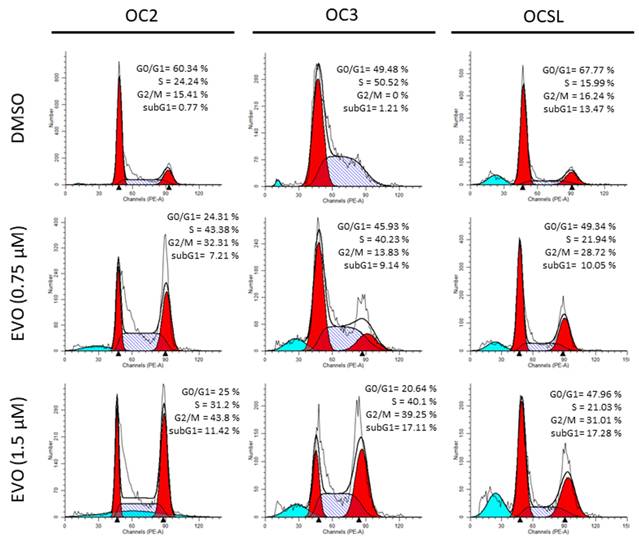
Considering that the growth inhibition of BN-OSCC cells under EVO treatment was evident, the cell cycle regulation of EVO treatment in OC2, OC3, and OCSL cells was verified. Figure 3 shows that the cell cycle progression of the abovementioned cells was mostly arrested at the G2/M phase. In addition, EVO slightly increased the subG1 population after 12 h (Fig. 3), indicating that EVO may exert a cytotoxic effect in these cells.
Evodiamine modulates cell cycle in BN-OSCC cells. Human BN-OSCC cell lines (OC2, OC3, and OCSL) were incubated with DMSO or EVO, and the cell cycle was examined with flow cytometry at 12 h post-incubation. DMSO was used as negative control.
Evodiamine induces cellular apoptosis in BN-OSCC cells
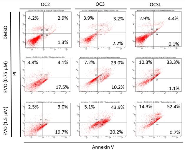
Considering that EVO administration can elevate the sub-G1 group in BN-OSCC cells (Fig. 3), cellular apoptosis induction in these cells was further evaluated using flow cytometry. The result showed that incubation with EVO could significantly elevate cellular apoptosis in BN-OSCC cells (Fig. 4). Moreover, EVO-mediated apoptosis in these cells was OC3 ≥ OCSL > OC2 cells (Fig. 4). These data indicated that EVO treatment induces cell cycle arrest and elevates cellular apoptosis in human BN-OSCC cells.
Evodiamine induces cellular apoptosis in human BN-OSCC cells. Human BN-OSCC cell lines (OC2, OC3, and OCSL) were administrated with evodiamine or DMSO for 24 h, and the apoptotic cells were examined with flow cytometry assay. DMSO was used as negative control.
Evodiamine induces caspase-dependent apoptosis in BN-OSCC cells
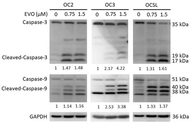
EVO induced apoptosis in a dose-dependent manner across all three BN-OSCC cell lines, as demonstrated by Annexin V/PI staining (Fig. 4). To further elucidate the underlying mechanism, the expression and cleavage of key apoptotic markers, including caspase-8, caspase-9, caspase-3, and poly (ADP-ribose) polymerase (PARP), were evaluated. The results showed the significant activation of caspase-9, caspase-3, and PARP following EVO treatment (Fig. 5), whereas caspase-8 remained unchanged. These findings indicate that EVO-induced apoptosis is primarily mediated through the intrinsic (mitochondrial) apoptotic pathway.
Evodiamine treatment elevates caspase activation in human BN-OSCC cells. Human BN-OSCC cell lines (OC2, OC3, and OCSL) were incubated with evodiamine or DMSO for 24 h, and the activation of caspases were examined with Western blot. DMSO was used as negative control. GAPDH was used as loading control.
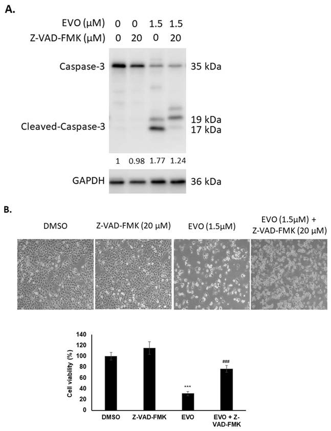
To further confirm the involvement of caspase-dependent apoptosis, the pan-caspase inhibitor Z-VAD-FMK was used to block EVO-induced cell death in OC3 cells. As shown in Fig. 6A, co-treatment with EVO and Z-VAD-FMK effectively attenuated caspase activation. Notably, cell viability was significantly restored in the combination treatment compared with EVO treatment alone (Fig. 6B). These results indicate that EVO-induced apoptosis in BN-OSCC cells is caspase-dependent.
Evodiamine treatment induces caspase-dependent apoptosis in human BN-OSCC cells. Human OC3 cells were incubated with Z-VAD-FMK, evodiamine or combination with Z-VAD-FMK and evodiamine for 24 h, and (A) the activation of caspases was examined with Western blot, and (B) the cell numbers or morphology of OC3 were determined with microscopy. DMSO was used as negative control. Z-VAD-FMK was used to block the activation of caspases. GAPDH was used as loading control. ***: comparing with DMSO group. ###: comparing with EVO group.
Discussion
OSCC is a heterogeneous malignancy associated with multiple etiological factors, including tobacco, alcohol consumption, and viral infections. By contrast, BN-OSCC is a distinct subtype primarily driven by chronic exposure to areca nut-related carcinogens [24]. Betel quid chewing remains a common habit in many regions of the world. Its major alkaloid component, arecoline, has been reported to trigger oxidative stress and genomic alterations, including DNA strand breaks and chromosomal instability, which are considered important events in oral carcinogenesis [25]. Compared with conventional OSCC, BN-OSCC exhibits unique biological characteristics, including increased oxidative stress, chronic inflammation, enhanced EMT, and a higher prevalence of genetic alterations, such as TP53 mutations [26-28]. Clinically, BN-OSCC is frequently associated with oral submucous fibrosis, which leads to trismus and significantly compromises surgical accessibility and margin clearance [29]. These differences contribute to several therapeutic challenges. Although the standard treatment modalities, including surgery, RT, and platinum-based chemotherapy, are similar for OSCC and BN-OSCC [30], patients with BN-OSCC often show reduced sensitivity to chemotherapy and RT, along with a higher risk of local recurrence caused by field cancerization [4, 31-34]. Furthermore, the fibrotic and chronically inflamed tumor microenvironment in BN-OSCC may impair drug delivery and modulate immune responses, resulting in variable outcomes with targeted therapy and immunotherapy [32-34]. Consequently, BN-OSCC is generally associated with poorer prognosis and greater clinical management difficulties than conventional OSCC. Therefore, the development of effective therapeutic approaches for BN-OSCC is urgently necessary.
EVO has positive effects on various cancers, including thyroid cancer, colorectal cancer, pancreatic cancer, gastric cancer, hepatoma, bladder cancer, tongue squamous cancer, oral cancer, and esophageal cancer [9, 14, 17, 35]. Although EVO has been shown to exert anti-OSCC activities, the effect of EVO on BN-OSCC remains unclear. In the present study, EVO exerted potent antitumor effects in human BN-OSCC cells, including the inhibition of cell proliferation, the induction of G2/M cell cycle arrest, and the activation of caspase-dependent apoptosis. Notably, EVO showed selective cytotoxicity toward BN-OSCC cell lines (OC2, OC3, and OCSL), whereas the non-betel nut-associated OSCC cell line (SAS) exhibited marked resistance (Fig. 1), indicating a distinct therapeutic vulnerability in BN-OSCC.
One of the most significant findings of this study is the differential sensitivity between BN-OSCC and non-BN-OSCC cells. The IC₅₀ values of EVO were only detectable in BN-OSCC cell lines, indicating a higher susceptibility to EVO treatment (Fig. 1). This observation is important because BN-OSCC exhibits more aggressive clinical behavior and therapeutic resistance than conventional OSCC [4, 31-34]. Given the selective inhibitory effect of EVO, it may target molecular pathways specifically dysregulated in BN-associated carcinogenesis, such as areca nut-induced oxidative stress, DNA damage, and aberrant signaling pathways [15, 32, 34, 36, 37]. However, differential responsiveness to EVO was observed among the BN-OSCC cell lines (Fig. 1). Furthermore, the timing and extent of EVO-induced apoptosis varied among the BN-OSCC cell lines (Fig. 4). A similar heterogeneity in drug responsiveness has been reported in oral cancer models. For example, OC2 and OCSL cells exhibit distinct proliferative, migratory, and invasive characteristics, as well as different sensitivities to piperlongumine treatment [38]. These findings indicate that the intrinsic biological heterogeneity among BN-OSCC cell lines may contribute to their differential responses to EVO. Nevertheless, the molecular mechanism underlying the relatively low sensitivity of OC2 cells to EVO remains unclear and warrants further investigation.
Mechanistically, our data indicate that EVO induces cell cycle arrest predominantly at the G2/M phase in BN-OSCC cells (Fig. 3). This finding is consistent with previous reports showing that EVO disrupts cell cycle progression by regulating key checkpoint proteins such as cyclin B1, CDK1, and p21 [9, 14, 39, 40]. G2/M arrest is often associated with DNA damage responses, and it serves as a critical checkpoint to prevent mitotic entry under cellular stress [41]. The observed increase in the sub-G1 population further indicates that prolonged cell cycle arrest may lead to apoptotic cell death (Figs. 3 and 4).
In addition to cell cycle arrest, EVO significantly induced apoptosis in BN-OSCC cells, as evidenced by Annexin V/PI staining and increased sub-G1 fraction (Figs. 3 and 4). Among the tested cell lines, OC3 and OCSL cells were more sensitive, followed by OC2 cells (Fig. 4), indicating potential heterogeneity in apoptotic responsiveness within BN-OSCC. Our mechanistic studies revealed that EVO-induced apoptosis is mediated through the intrinsic (mitochondrial) pathway, as demonstrated by the activation of caspase-9, caspase-3, and PARP cleavage, whereas caspase-8 remained unaffected (Fig. 5). Therefore, EVO primarily triggers mitochondrial dysfunction rather than death receptor-mediated apoptosis.
Furthermore, the use of the pan-caspase inhibitor Z-VAD-FMK significantly attenuated EVO-induced caspase activation and restored cell viability, confirming that apoptosis induced by EVO is caspase-dependent (Fig. 6). These findings are consistent with previous studies on other cancer types [9, 42-44], where EVO induces mitochondrial apoptosis by modulating Bcl-2 family proteins, reactive oxygen species generation, and the loss of mitochondrial membrane potential.
From a clinical perspective, the selective antitumor activity of EVO against BN-OSCC cells is particularly noteworthy. Patients with BN-OSCC often have a poorer prognosis and limited therapeutic options because of intrinsic or acquired resistance to conventional chemotherapy and RT. The ability of EVO to selectively target BN-OSCC cells while sparing non-BN-OSCC cells indicates its potential application as a novel therapeutic agent or adjuvant treatment for this specific subtype of oral cancer.
This study has several limitations that should be acknowledged. First, this study was conducted primarily in vitro using established cell lines, which may not fully recapitulate the tumor microenvironment in vivo. Second, the molecular targets underlying the selective sensitivity of BN-OSCC to EVO remain unclear and warrant further investigation. Thus, future studies should identify the key signaling pathways involved and validate these findings in animal models and clinical samples.
In conclusion, our study provides evidence that EVO selectively inhibits proliferation and induces caspase-dependent apoptosis in BN-OSCC cells through G2/M cell cycle arrest and the activation of the intrinsic apoptotic pathway. These findings highlight the potential application of EVO as a promising therapeutic agent for BN-OSCC and support further preclinical and clinical investigations.
Acknowledgements
Funding
This work was supported by grants from Taiwan's National Science and Technology Council (NSTC 114-2314-B-037-077-MY3), Ministry of Agriculture (115AS-1.3.2-AS-04), Ditmanson Medical Foundation Chia-Yi Christian Hospital (R114-32), and Kaohsiung Medical University Research Center, Kaohsiung Medical University (KMU-TC114B01).
Author contributions
The authors confirm contribution to the paper as follows: Conceptualization, Ying-Ray Lee, Yin-Che Lu, and Chin-Ho Kuo; methodology, Ying-Ray Lee, Kai-Liang Tang, Chih-Hsien Liao, and Yi-Zhen Li; validation, Chih-Hsien Liao, Yi-Zhen Li, Pin-Tzu Chen, and Shu-Hsin Chen; formal analysis, Chih-Hsien Liao, Yi-Zhen Li, Pin-Tzu Chen, and Shu-Hsin Chen; investigation, Chih-Hsien Liao, and Yi-Zhen Li; data curation, Chih-Hsien Liao, Yi-Zhen Li, and Shu-Hsin Chen; writing-original draft preparation, Ying-Ray Lee; writing-review and editing, Ying-Ray Lee, Yin-Che Lu, and Kai-Liang Tang; visualization, Chih-Hsien Liao, and Yi-Zhen Li; supervision, Ying-Ray Lee; project administration, Ying-Ray Lee; funding acquisition, Ying-Ray Lee and Yin-Che Lu, and Kai-Liang Tang. All authors reviewed the results and approved the final version of the manuscript.
Declaration of generative AI use
During the preparation of this work the authors used ChatGPT in order to assist in tasks such as language polishing and improving readability. After using this tool, the authors reviewed and edited the content as needed and takes full responsibility for the content of the published article.
Competing Interests
The authors have declared that no competing interest exists.
References
1. Siegel R, Naishadham D, Jemal A. Cancer statistics, 2013. CA Cancer J Clin. 2013;63:11-30
2. Chung TT, Pan MS, Kuo CL, Wong RH, Lin CW, Chen MK. et al. Impact of RECK gene polymorphisms and environmental factors on oral cancer susceptibility and clinicopathologic characteristics in Taiwan. Carcinogenesis. 2011;32:1063-8
3. Jemal A, Bray F, Center MM, Ferlay J, Ward E, Forman D. Global cancer statistics. CA Cancer J Clin. 2011;61:69-90
4. Liao CT, Wallace CG, Lee LY, Hsueh C, Lin CY, Fan KH. et al. Clinical evidence of field cancerization in patients with oral cavity cancer in a betel quid chewing area. Oral Oncol. 2014;50:721-31
5. Trivedy CR, Craig G, Warnakulasuriya S. The oral health consequences of chewing areca nut. Addict Biol. 2002;7:115-25
6. Lee YR, Wu WC, Ji WT, Chen JY, Cheng YP, Chiang MK. et al. Reversine suppresses oral squamous cell carcinoma via cell cycle arrest and concomitantly apoptosis and autophagy. J Biomed Sci. 2012;19:9
7. Pointreau Y, Garaud P, Chapet S, Sire C, Tuchais C, Tortochaux J. et al. Randomized trial of induction chemotherapy with cisplatin and 5-fluorouracil with or without docetaxel for larynx preservation. J Natl Cancer Inst. 2009;101:498-506
8. Andreadis C, Vahtsevanos K, Sidiras T, Thomaidis I, Antoniadis K, Mouratidou D. 5-Fluorouracil and cisplatin in the treatment of advanced oral cancer. Oral Oncol. 2003;39:380-5
9. Shi CS, Li JM, Chin CC, Kuo YH, Lee YR, Huang YC. Evodiamine Induces Cell Growth Arrest, Apoptosis and Suppresses Tumorigenesis in Human Urothelial Cell Carcinoma Cells. Anticancer Res. 2017;37:1149-59
10. Yu H, Jin H, Gong W, Wang Z, Liang H. Pharmacological actions of multi-target-directed evodiamine. Molecules. 2013;18:1826-43
11. Lin L, Liu Y, Tang R, Ding S, Lin H, Li H. Evodiamine: A Extremely Potential Drug Development Candidate of Alkaloids from Evodia rutaecarpa. Int J Nanomedicine. 2024;19:9843-70
12. Shoji N, Umeyama A, Takemoto T, Kajiwara A, Ohizumi Y. Isolation of evodiamine, a powerful cardiotonic principle, from Evodia rutaecarpa Bentham (Rutaceae). J Pharm Sci. 1986;75:612-3
13. Panda M, Tripathi SK, Zengin G, Biswal BK. Evodiamine as an anticancer agent: a comprehensive review on its therapeutic application, pharmacokinetic, toxicity, and metabolism in various cancers. Cell Biol Toxicol. 2023;39:1-31
14. Yu HI, Chang HY, Lu CH, Tai TS, Kung FP, Zhang YS. et al. Evodiamine exerts anti-cancer activity including growth inhibition, cell cycle arrest, and apoptosis induction in human follicular thyroid cancers. Am J Cancer Res. 2024;14:4989-99
15. Yu HI, Chou HC, Su YC, Lin LH, Lu CH, Chuang HH. et al. Proteomic analysis of evodiamine-induced cytotoxicity in thyroid cancer cells. J Pharm Biomed Anal. 2018;160:344-50
16. Solanki R, Patel S. Evodiamine and its nano-based approaches for enhanced cancer therapy: recent advances and challenges. J Sci Food Agric. 2024;104:8430-44
17. Lu YC, Lin TH, Tang KL, Kuo CH, Zhang YS, Chang YP. et al. The impact of evodiamine on human anaplastic thyroid cancer therapy-an in vitro and in vivo study. Int J Med Sci. 2025;22:4532-46
18. Sachita K, Kim Y, Yu HJ, Cho SD, Lee JS. In Vitro Assessment of the Anticancer Potential of Evodiamine in Human Oral Cancer Cell Lines. Phytother Res. 2015;29:1145-51
19. Wu Y, Wang J, Zhao J, Zhang Y, Sun Y, Chen J. et al. Gene regulation analysis of the effects of evodiamine on tongue squamous cell carcinoma. J Cell Biochem. 2019;120:15933-40
20. Ren L, Lou Y, Sun M. The anti-tumor effects of evodiamine on oral squamous cell carcinoma (OSCC) through regulating advanced glycation end products (AGE) / receptor for advanced glycation end products (RAGE) pathway. Bioengineered. 2021;12:5985-95
21. Lin LQ, Lv SY, Ren HZ, Li RR, Li L, Pang YQ. et al. Evodiamine inhibits EPRS expression to regulate glutamate metabolism and proliferation of oral squamous cell carcinoma cells. Kaohsiung J Med Sci. 2024;40:348-59
22. Wu JJ, Chen SH, Lee CH, Li YZ, Hsu YW, Hsieh MY. et al. Flavopereirine exerts anti-cancer activities in various human thyroid cancer cells. Am J Cancer Res. 2024;14:3317-34
23. Liao KS, Lee YR, Chao WY, Huang YJ, Chung HC, Chen SH. et al. Honokiol Suppresses Cell Proliferation and Tumor Migration through ROS in Human Anaplastic Thyroid Cancer Cells. Endocr Metab Immune Disord Drug Targets. 2024
24. Warnakulasuriya S. Global epidemiology of oral and oropharyngeal cancer. Oral Oncol. 2009;45:309-16
25. Wang WC, Chiu YT, Wang YY, Lu SL, Chan LP, Lee CY. et al. Effects of DSM-5 Betel-Quid-Related Symptoms, Pathological Behaviors, and Use Disorder on Oral Squamous Cell Carcinoma Risk. Cancers (Basel). 2022 14
26. Senevirathna K, Pradeep R, Jayasinghe YA, Jayawickrama SM, Illeperuma R, Warnakulasuriya S. et al. Carcinogenic Effects of Areca Nut and Its Metabolites: A Review of the Experimental Evidence. Clin Pract. 2023;13:326-46
27. Chen IH, Chang JT, Liao CT, Wang HM, Hsieh LL, Cheng AJ. Prognostic significance of EGFR and Her-2 in oral cavity cancer in betel quid prevalent area cancer prognosis. Br J Cancer. 2003;89:681-6
28. Takeshima M, Saitoh M, Kusano K, Nagayasu H, Kurashige Y, Malsantha M. et al. High frequency of hypermethylation of p14, p15 and p16 in oral pre-cancerous lesions associated with betel-quid chewing in Sri Lanka. J Oral Pathol Med. 2008;37:475-9
29. Lee YH, Yang LC, Hu FW, Peng CY, Yu CH, Yu CC. Elevation of Twist expression by arecoline contributes to the pathogenesis of oral submucous fibrosis. J Formos Med Assoc. 2016;115:311-7
30. Colevas AD, Yom SS, Pfister DG, Spencer S, Adelstein D, Adkins D. et al. NCCN Guidelines Insights: Head and Neck Cancers, Version 1.2018. J Natl Compr Canc Netw. 2018;16:479-90
31. Yang J, Wang ZY, Huang L, Yu TL, Wan SQ, Song J. et al. Do betel quid and areca nut chewing deteriorate prognosis of oral cancer? A systematic review, meta-analysis, and research agenda. Oral Dis. 2021;27:1366-75
32. Zhang T, Xiong H, Zeng L, Yang Z, Hu X, Su T. The key target and role of betel nut in oral squamous cell carcinoma. BMC Oral Health. 2025;25:1212
33. Sreekissoon S, Wang D, Wang Y, Wang C, Neda TS, Zhu M. et al. Distinctive tumor biology of oral squamous cell carcinoma associated with oral submucous fibrosis and its suppressive immune microenvironment. BMC Oral Health. 2025;26:347
34. Su SC, Lin CW, Chen MK, Lee YC, Su CW, Bai S. et al. Multimodal profiling of oral squamous cell carcinoma identifies genomic alterations and expression programs associated with betel quid chewing. Neoplasia. 2025;68:101218
35. Zhou Z, Zhou Y, Zhang Z, Zhao M, Hu C, Yang L. et al. Progress on the effects and underlying mechanisms of evodiamine in digestive system diseases, and its toxicity: A systematic review and meta-analysis. Phytomedicine. 2024;132:155851
36. Li B, Liu J, Zhang D, Chu Y, Chen Z, Tsao J. et al. Evodiamine Promotes Autophagy and Alleviates Oxidative Stress in Dry Eye Disease Through the p53/mTOR Pathway. Invest Ophthalmol Vis Sci. 2025;66:44
37. Yang J, Wu LJ, Tashiro S, Onodera S, Ikejima T. Nitric oxide activated by p38 and NF-kappaB facilitates apoptosis and cell cycle arrest under oxidative stress in evodiamine-treated human melanoma A375-S2 cells. Free Radic Res. 2008;42:1-11
38. Chen SY, Liu GH, Chao WY, Shi CS, Lin CY, Lim YP. et al. Piperlongumine Suppresses Proliferation of Human Oral Squamous Cell Carcinoma through Cell Cycle Arrest, Apoptosis and Senescence. Int J Mol Sci. 2016 17
39. Zhong ZF, Tan W, Wang SP, Qiang WA, Wang YT. Anti-proliferative activity and cell cycle arrest induced by evodiamine on paclitaxel-sensitive and -resistant human ovarian cancer cells. Sci Rep. 2015;5:16415
40. Fang C, Zhang J, Qi D, Fan X, Luo J, Liu L. et al. Evodiamine induces G2/M arrest and apoptosis via mitochondrial and endoplasmic reticulum pathways in H446 and H1688 human small-cell lung cancer cells. PLoS One. 2014;9:e115204
41. Ree AH, Stokke T, Bratland A, Patzke S, Nome RV, Folkvord S. et al. DNA damage responses in cell cycle G2 phase and mitosis-tracking and targeting. Anticancer Res. 2006;26:1909-16
42. Liu L, Sun X, Guo Y, Ge K. Evodiamine induces ROS-Dependent cytotoxicity in human gastric cancer cells via TRPV1/Ca(2+) pathway. Chem Biol Interact. 2022;351:109756
43. Chien CC, Wu MS, Chou SW, Jargalsaikhan G, Chen YC. Roles of reactive oxygen species, mitochondrial membrane potential, and p53 in evodiamine-induced apoptosis and G2/M arrest of human anaplastic thyroid carcinoma cells. Chin Med. 2021;16:134
44. Fang Q, Jiang S, Li C. Evodiamine Selectively Inhibits Multiple Myeloma Cell Growth by Triggering Activation of Intrinsic Apoptosis Pathway. Onco Targets Ther. 2019;12:11383-91
Author contact
Corresponding author: Ying-Ray Lee, Ph. D., Department of Microbiology and Immunology, College of Medicine, Kaohsiung Medical University, Kaohsiung, Taiwan, No. 100, Shih-Chuan 1st Rd., Kaohsiung City 80708, Taiwan, Tel: +886-7-3121101 Ext.2150#12, E-mail: yingray.leecom.