Impact Factor
ISSN: 1837-9664
J Cancer 2017; 8(10):1795-1800. doi:10.7150/jca.18848 This issue Cite
Research Paper
PART1 expression is associated with poor prognosis and tumor recurrence in stage I-III non-small cell lung cancer
1. Department of Respiratory Medicine, Shanghai 10th People's Hospital, Tongji University, Shanghai 200072, China.
2. Department of Endocrinology, Xinhua Hospital, Shanghai Jiaotong University School of Medicine, Shanghai 200092, China
3. Institute of Biochemistry and Biology, Shanghai Institutes for Biological Sciences, Chinese Academy of Sciences, Shanghai 200031, China.
*These authors have contributed equally to this work.
Received 2016-12-21; Accepted 2017-4-26; Published 2017-7-1
Abstract
Purpose: Long noncoding RNAs (lncRNAs) have been demonstrated as effective markers for cancer detection and prognosis prediction. The aim of this study was to determine whether tissue PART1 could be used as a biomarker for prognosis prediction of non-small cell lung cancer (NSCLC).
Methods: PART1 expression was detected in 208 cases of stage I-III NSCLC specimens and adjacent normal tissues. The Cox proportional-hazards regression model was used to analyze the association between PART1 expression and overall survival (OS) and disease-free survival (DFS) of the patients.
Results: It was found that the expression of PART1 was significantly up-regulated in 73.1% (152/208) stage I-III NSCLC specimens compared with adjacent normal tissues. High tissue PART1 expression was associated with shorter OS and could serve as an independent prognostic biomarker in stage I-III NSCLC patients (hazard ratio [HR] = 2.11, 95% CI = 1.18-3.78, P = 0.012). In addition, high tissue PART1 expression indicated poor DFS in stage I-III NSCLC patients (HR = 1.94, 95% CI: 1.37-2.76, P < 0.001).
Conclusions: PART1 may prove to be a promising biomarker for prediction of survival and tumor recurrence in stage I-III NSCLC.
Keywords: non-small-cell lung carcinoma, lncRNAs, prostate androgen-regulated transcript-1, prognosis, biomarkers.
Introduction
Lung cancer is one of the most common human cancers, and the leading cause of cancer-related mortality, resulting in an estimated 224,210 new diagnoses and 159,260 deaths in the United States in 2014 [1]. Non-small cell lung cancer (NSCLC) represents approximately 85% of all lung cancers, and is often discovered at the locally advanced or metastatic stage of the disease. Despite recent advances in targeted therapies, the median 5-year survival rate of NSCLC patients is only about 5% worldwide [2]. Therefore, prognostic assessment of NSCLC patients is essential for the choice of better therapeutic strategies. Although previous studies have identified various differentially expressed protein-coding genes in NSCLC, they lack sufficient sensitivity and specificity, especially for early diagnosis, prognosis assessment and prediction of recurrence, and therefore novel molecular markers are urgently needed for clinical use [3].
Recently, growing evidence suggests that long non-coding RNAs (lncRNAs) similar to protein-coding genes can act as either oncogenes or tumor suppressors and may be a new class of cancer biomarkers and therapeutic targets [4]. MALAT1 (metastasis-associated lung adenocarcinoma transcript 1) is known as a key prognostic biomarker for lung adenocarcinoma [5]. HOTAIR (HOX antisense intergenic RNA) is reported to be highly expressed in NSCLC with advanced stage and lymph node metastasis [6]. Low expression of taurine upregulated gene 1 (TUG1) [7] and BRAF activated noncoding RNA (BANCR) [8] are associated with larger tumor size, advanced pathological stage and poorer overall survival (OS). Prostate androgen regulated transcript 1 (PART1) is known as a human prostate-specific and androgen-regulated gene that maps to chromosome 5q12. It is highly expressed in the prostate gland and plays an important role in prostate carcinogenesis [9]. However, studies on the role of PART1 in human tumors are still limited, and very little is known about the clinical utility and accuracy of PART1 in NSCLC.
In the present study, we investigated the expression of lncRNA PART1 in clinical NSCLC specimens and adjacent normal tissues, in an attempt to clarify its association with OS and disease-free survival (DFS) of NSCLC patients.
Results
PART1 expression in NSCLC specimens and adjacent normal tissues
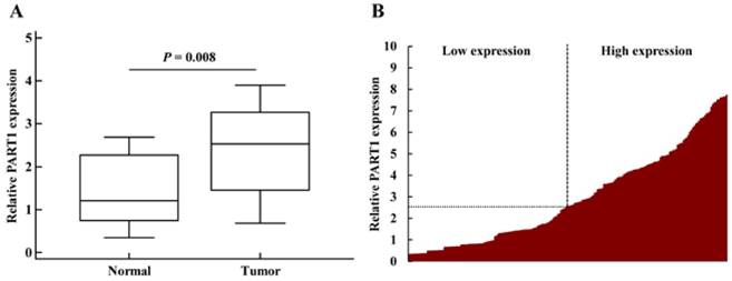
In the initial screening phase, a total of 20 paired stage I-III NSCLC specimens and adjacent normal tissues were analyzed using qRT-PCR. It was found that the expression of PART1 was significantly up-regulated in stage I-III NSCLC specimens compared with adjacent normal tissues (P = 0.008) (Figure 1A). To validate whether PART1 was differentially expressed in cancer tissues, the relative expression of PART1 in stage I-III NSCLC specimens (n = 208) in comparison with corresponding non-tumor normal tissues was examined by qRT-PCR. PART1 was detected in all 208 (100%) NSCLC tissues. As shown in Figure 1B, PART1 expression was significantly up-regulated in 73.1% (152/208) NSCLC specimens compared with adjacent normal tissues. The above results suggest that over-expression of PART1 might be involved in the pathogenesis of NSCLC.
Association between PART1 expression and clinicopathologic characteristics of patients
Subsequently, we analyzed the correlation between PART1 expression and the clinicopathologic characteristics of the NSCLC patients. According to the median level of PART1 (fold change = 2.5), the patients were assigned to two groups: PART1 high expression group (n= 104) and PART1 low expression group (n = 104). As shown in Table 1, high PART1 expression was more frequently observed in squamous tumors (P = 0.037). However, there was no significant correlation between PART1 expression and the smoking status (P = 0.405), tumor differentiation (P = 0.867) or epidermal growth factor receptor (EGFR) mutations (P = 0.256).
Association between PART1 expression in tissue and patients' characteristics.
| Factors | Tissue PART1 (n = 208) | P | |
|---|---|---|---|
| Low (n = 104) | High (n = 104) | ||
| Age (years) | 0.089 | ||
| ≤ 65 | 48 | 35 | |
| > 65 | 56 | 69 | |
| Gender | 0.127 | ||
| Male | 45 | 57 | |
| Female | 59 | 47 | |
| Smoking status | 0.405 | ||
| Nonsmoker | 48 | 55 | |
| Ever-smoker | 56 | 49 | |
| Histologic type | 0.037 | ||
| Squamous | 39 | 55 | |
| Non-squamous | 65 | 49 | |
| T status | 0.694 | ||
| T1-2 | 62 | 67 | |
| T3-4 | 42 | 39 | |
| N status | 0.553 | ||
| N0 | 68 | 73 | |
| N1-3 | 36 | 31 | |
| Stage | 0.774 | ||
| I-II | 64 | 67 | |
| III | 40 | 37 | |
| Differentiation | 0.867 | ||
| well, moderate | 80 | 82 | |
| poor | 24 | 22 | |
| EGFR mutation status | 0.256 | ||
| Mutated | 21 | 29 | |
| Wild type | 83 | 75 | |
Correlation between PART1 expression and survival of NSCLC patients
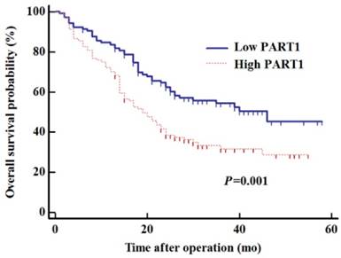
To determine whether tissue PART1 levels could predict NSCLC prognosis, survival analysis was performed to estimate OS of NSCLC patients. Kaplan-Meier analysis demonstrated that patients with high PART1 expression in cancer tissues had poorer survival than those with low PART1 expression (Figure 2, log-rank test: P = 0.001). To determine whether PART1 expression was an independent risk factor for prognosis, the Cox proportional-hazards regression was used for survival data (Table 2). Univariate regression analysis showed that high tissue level of PART1 (P = 0.002), high T stage (T3/4; P < 0.001), lymph node metastasis (P = 0.002), and poor differentiation (P = 0.030) were significantly correlated with poor OS (Table 2). Furthermore, multivariate analysis revealed that high PART1 expression was a significant prognostic factor for poorer OS (HR = 2.11, 95% CI = 1.18-3.78, P = 0.012), independent of T stage (HR = 2.36, 95% CI: 1.54- 3.61, P < 0.001, Table 2). The above results indicate that high tissue PART1 expression was an independent marker for poor OS in stage I-III NSCLC patients.
Correlation between PART1 expression and recurrence risk of NSCLC patients
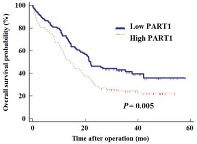
To further evaluate whether tissue PART1 level could serve as a predictor of tumor recurrence, the Cox proportional-hazards regression was employed. Similar to the result on OS, there was a significant correlation between DFS and PART1 expression level (Figure 3, log-rank test: P = 0.005). Univariate regression analysis also demonstrated that high T stage (T3/4, P = 0.021), lymph node metastasis (P = 0.009), and poor differentiation (P = 0.042) were significantly correlated with DFS. Furthermore, multivariate analysis showed that high PART1 expression was a significant predictor for tumor recurrence (P < 0.001, HR = 1.94, 95% CI: 1.37-2.76; Table 3), independent of T stage (HR = 1.68, 95% CI: 1.13-2.49, P = 0.011) and differentiation (HR = 1.50, 95% CI: 1.02-2.22, P = 0.043, Table 3). These results indicate that stage I-III NSCLC patients with high PART1 expression had a higher risk of recurrence than those with low PART1 expression.
Discussion
The present study investigated the potential clinical utility of lncRNA PART1 as a novel prognostic and metastasis-predictive biomarker in patients with stage I-III NSCLC. We found that the expression of PART1 was significantly up-regulated in most NSCLC specimens. Furthermore, stage I-III NSCLC patients with high PART1 expression had a shorter OS and a higher risk of recurrence than those with low PART1 expression.
Relative expression of PART1 in tumor and normal tissues. A: Expression of PART1 in 20 paired stage I-III NSCLC specimens and adjacent normal tissues. The boxes represent the interquartile range, and the lines across the boxes indicate the median values. B: NSCLC patients were devided into two groups: high PART1 group (fold change≥2.5) and low PART1 group (fold change <2.5). Data were presented as relative expression of PART1 in NSCLC tissues normalized to adjacent normal tissues. All the experiments were performed 3 times independently.

Univariate and multivariate analyses for prognostic factors in patients with NSCLC.
| Variables | Univariate | Multivariate | |||||
|---|---|---|---|---|---|---|---|
| HR | 95% CI | P | HR | 95% CI | P | ||
| Age, years (> 65 vs ≤ 65) | 0.80 | 0.55-1.18 | 0.245 | — | — | — | |
| Gender (female vs male) | 1.17 | 0.81-1.68 | 0.393 | — | — | — | |
| Smoking (No vs Yes) | 1.08 | 0.75-1.55 | 0.674 | — | — | — | |
| Histology (squamous vs non-squamous) | 1.19 | 0.83-1.71 | 0.351 | — | — | — | |
| Pathological T (T3-4 vs T1-2) | 1.95 | 1.28-2.98 | <0.001 | 2.36 | 1.54-3.61 | <0.001 | |
| Lymph node metastasis (N1-3 vs N0) | 1.75 | 1.18-2.61 | 0.002 | 1.46 | 0.99-2.15 | 0.059 | |
| Differentiation (poor vs well/moderate) | 1.56 | 0.98-2.51 | 0.030 | 1.48 | 0.97-2.27 | 0.070 | |
| EGFR mutation status (No vs Yes) | 1.31 | 0.85-2.04 | 0.182 | — | — | — | |
| PART1 in tumor (high vs low) | 1.80 | 1.25-2.60 | 0.001 | 2.11 | 1.18-3.78 | 0.012 | |
HR, hazard ratio; CI, confidence interval.
Univariate and multivariate analyses for predictive factors of recurrence in NSCLC patients.
| Variables | Univariate | Multivariate | |||||
|---|---|---|---|---|---|---|---|
| HR | 95% CI | P | HR | 95% CI | P | ||
| Age, years (> 65 vs ≤ 65) | 0.86 | 0.61-1.21 | 0.387 | — | — | — | |
| Gender (female vs male) | 1.24 | 0.89-1.73 | 0.197 | — | — | — | |
| Smoking (No vs Yes) | 1.29 | 0.93-1.80 | 0.126 | — | — | — | |
| Histology (squamous vs non-squamous) | 1.23 | 0.88-1.72 | 0.218 | — | — | — | |
| Pathological T (T3-4 vs T1-2) | 1.52 | 1.07-2.17 | 0.021 | 1.68 | 1.13-2.49 | 0.011 | |
| Lymph node metastasis (N1-3 vs N0) | 1.57 | 1.12-2.20 | 0.009 | 1.43 | 1.00-2.03 | 0.051 | |
| Differentiation (poor vs well/moderate) | 1.49 | 1.02-2.20 | 0.042 | 1.50 | 1.02-2.22 | 0.043 | |
| EGFR mutation status (No vs Yes) | 0.77 | 0.53-1.13 | 0.183 | — | — | — | |
| PART1 in tumor (high vs low) | 1.61 | 1.15-2.24 | 0.005 | 1.94 | 1.37-2.76 | <0.001 | |
HR, hazard ratio; CI, confidence interval.
Kaplan-Meier estimates of the overall survival in NSCLC patients subdivided by tissue PART1 levels.

Kaplan-Meier estimates of the disease-free survival in NSCLC patients subdivided by tissue PART1 levels.

With the development of microarray technology, lncRNAs have become an attractive focus in the field of genetic science. lncRNAs have been observed in various human diseases including cancers. Experimental studies showed that several lncRNAs were deregulated in tumor tissues of various cancer types. For example, Ji et al [10] identified MALAT1 as a prognostic marker for metastasis and patient survival in NSCLC. In addition, MALAT1 might also affect the growth and colony formation of NSCLC cells in vitro [11]. Liu et al [12] showed that HOTAIR regulated NSCLC cell invasion, and that high level of HOTAIR expression indicated a poor prognosis in NSCLC. Cheng et al [13] reported that over-expression of UCA1 (urothelial cancer-associated 1) was significantly associated with a shorter progression-free survival in NSCLC. Furthermore, the deregulation of lncRNAs in primary tumor tissues was mirrored in plasma and urine. For instance, Weber et al [14] found that MALAT1 was a candidate blood-based biomarker for the diagnosis of NSCLC. Zhou et al [15] reported that plasma H19 could serve as a potential biomarker for the diagnosis of gastric cancer. Wang et al [16] demonstrated that urine UCA1 was a very sensitive and specific unique marker for bladder cancer. However, we did not restrict our research to plasma PART1 because of the minute quantity and unstable expression of RNA isolated from blood. As mentioned previously, PART1 expression was significantly up-regulated in most NSCLC specimens. Therefore, we conducted this study and proved the relationship between tissue PART1 expression and prognosis in stage I-III NSCLC.
PART-1 is a long non-coding RNA located on chromosome 5q12. Previous studies focused on prostate gland disease. Lin et al [9] demonstrated that PART-1 was highly expressed in the prostate gland and regulated by androgens in human prostate cancer cells. Sidiropoulos et al [17] found that PART-1 was more frequently overexpressed in the cancerous prostatic tissue than that in the benign tissue. Yu et al [18] reported that PART-1 expression was upregulated by androgens and the upregulation was significantly downregulated by genistein in androgen-sensitive LNCaP cells. A recent study [19] identified a set of six lncRNAs (including PART1) that were significantly associated with OS in glioblastoma multiforme. Hou et al [20] performed a genome-wide gene expression analysis on a cohort of 91 NSCLC patients. Re-analysis of the data showed that PART1 was overexpressed in squamous cell lung cancer tissue as compared with that in the normal lung tissue. Our study further confirmed that PART1 expression was up-regulated in most NSCLC specimens, and high PART1 expression predicted a shorter OS and a higher risk of recurrence. Therefore, the present study could serve as a foundation for future study on the role of PART-1 as a lung cancer marker.
The study has several strengths including its prospective design, large sample size, completeness of follow-up and use of multivariable adjustments. We restricted the inclusion of patients who had not received neoadjuvant chemotherapy to avoid possible influence on PART1 expression. In addition, we also limited our inclusion to patients with stage I-III NSCLC who received surgical treatment. Therefore, sufficient NSCLC specimens and adjacent normal tissues were curtained. After that, we examined the relative expression of PART1 in NSCLC specimens in comparison with corresponding non-tumor normal tissues to minimize individual differences.
This study has some limitations, including the limited clinical information and high heterogeneity in postoperative chemotherapy regimens. In addition, the possibility of patient selection bias could not be excluded, because some patients were unable to tolerate surgery due to comorbidities, terminal illness, or poor performance status. As our conclusion is limited to stage I-III NSCLC patients, further research is needed to confirm our conclusion in all stage NSCLC patients.
In summary, our study provides evidence that PART1 is an independent prognostic factor of OS and DFS for stage I-III NSCLC patients. Future study should focus on the verification of our findings and the biological function of PART1 in lung cancer tissues.
Materials and Methods
Study Design
The present research was approved by the Ethics Committees of Shanghai 10th People's Hospital and Shanghai Pulmonary Disease Hospital (Shanghai, China). All patients or family members provided written informed consent and indicated willingness to donate their tissue samples for research. The inclusion criteria were patients who were histologically diagnosed with NSCLC by two senior pathologists, provided sufficient NSCLC specimens and normal tissues for RNA isolation, and received complete tumor resection. The exclusion criteria were patients who had received chemotherapy or radiotherapy before surgery; patients with a self-reported previous history of cancer history or who were diagnosed with new cancers; patients with metastasis from other organs, and those who refused to sign the informed consent.
Briefly, a total of 208 NSCLC patients from Shanghai Pulmonary Disease Hospital (Shanghai, China) between January 2009 and May 2009 were enrolled in this study. Fresh clinical NSCLC specimens and adjacent normal tissues were collected within 10 minutes after surgical removal. The samples were then dipped into liquid nitrogen and conserved in a -80°C freezer for mRNA extraction. Clinicopathologic features and follow-up data of the 208 patients were collected at the first visit and follow-ups every 3 month by telephone.
The present study was designed as a screening phase and a validation phase. In the initial screening phase, tissue-specific expression of PART1 was analyzed in a small group of 20 NSCLC patients. In the subsequent validation phase, PART1 expression in tissues from NSCLC patients were evaluated in a large and independent cohort of 208 patients.
RNA isolation and qRT-PCR from tissues
Total RNA was extracted from NSCLC specimens and adjacent normal tissues using Trizol reagent (Invitrogen, Carlsbad, CA) according to the manufacturer's protocol. Briefly, the sample was homogenized in 10 volume TRI Reagent and centrifuged at 12,000 xg for 10 min at 4°C. The supernatant was transferred to a fresh tube, to which 100 μL BCP per 1 mL of TRI Reagent solution was added for homogenization. The tube was then centrifuged at 7,500 x g for 5 min to remove the ethanol. The RNA pellet was air dried briefly. Total RNA was dissolved in THE RNA Storage Solution. cDNA for RT-PCR was synthesized with the PrimeScript RT Master Mix (Perfect Real Time) kit. β-actin was used as an internal control. PART1 primers used were: Forward 5'-AAGGCCGTGTCAGAACTCAA-3' and Reverse 5'-GTTTTCCATCTCAGCCTGGA-3'. β-actin primers used were: Forward 5'-ATCATGTTTGAGACCTTCAACA-3' and Reverse 5'-CATCTCTTGCTCGAAGTCCA-3'. qRT-PCR was performed in an ABI Prism 7600 system (Applied Biosystems, USA) according to the manufacturer's instructions, with the following cycling conditions: 95℃ for 10 min, followed by 45 cycles at 95℃ for 15s and 60℃ for 45 s. The gene expression data was calculated with the 2-ΔΔCt method.
Statistical Analysis
Statistical analysis was carried out using Medcalc version 11.2 (Broekstraat52, Mariakerke, Belgium). χ2 test was used for categoric data. Survival curves were estimated using the Kaplan-Meier method, and differences in survival distributions were evaluated by the log-rank test. OS and DFS were measured for each patient. The Cox proportional-hazards regression model was used to estimate hazard ratios for recurrence and prognosis. A value of < 0.05 was considered statistically significant.
Acknowledgements
This work was supported by Science and Technology Commission of Shanghai Municipality (14411971200) and the National Natural Science Foundation of China (81372175 and 81172229). The funders had no role in study design, data collection and analysis, decision to publish, or preparation of the manuscript.
Ethical approval
The study was reviewed and approved by the Institutional Ethics Committee of the Shanghai Tenth People's Hospital of Tongji University and was conducted in compliance with the Helsinki Declaration. Written informed consent was obtained from all subjects.
Competing Interests
The authors have declared that no competing interests exist.
References
1. Siegel R, Desantis C, Jemal A. Colorectal cancer statistics, 2014. CA: a cancer journal for clinicians. 2014;64(2):104-117
2. Jemal A, Siegel R, Ward E, Hao Y, Xu J, Thun MJ. Cancer statistics, 2009. CA: a cancer journal for clinicians. 2009;59(4):225-249
3. Cheetham SW, Gruhl F, Mattick JS, Dinger ME. Long noncoding RNAs and the genetics of cancer. British journal of cancer. 2013;108(12):2419-2425
4. Kung JT, Colognori D, Lee JT. Long noncoding RNAs: past, present, and future. Genetics. 2013;193(3):651-669
5. Gutschner T, Hammerle M, Eissmann M, Hsu J, Kim Y, Hung G, Revenko A, Arun G, Stentrup M, Gross M, Zornig M, MacLeod AR, Spector DL, Diederichs S. The noncoding RNA MALAT1 is a critical regulator of the metastasis phenotype of lung cancer cells. Cancer research. 2013;73(3):1180-1189
6. Nakagawa T, Endo H, Yokoyama M, Abe J, Tamai K, Tanaka N, Sato I, Takahashi S, Kondo T, Satoh K. Large noncoding RNA HOTAIR enhances aggressive biological behavior and is associated with short disease-free survival in human non-small cell lung cancer. Biochemical and biophysical research communications. 2013;436(2):319-324
7. Zhang EB, Yin DD, Sun M, Kong R, Liu XH, You LH, Han L, Xia R, Wang KM, Yang JS, De W, Shu YQ, Wang ZX. P53-regulated long non-coding RNA TUG1 affects cell proliferation in human non-small cell lung cancer, partly through epigenetically regulating HOXB7 expression. Cell death & disease. 2014;5:e1243
8. Sun M, Liu XH, Wang KM, Nie FQ, Kong R, Yang JS, Xia R, Xu TP, Jin FY, Liu ZJ, Chen JF, Zhang EB, De W, Wang ZX. Downregulation of BRAF activated non-coding RNA is associated with poor prognosis for non-small cell lung cancer and promotes metastasis by affecting epithelial-mesenchymal transition. Molecular cancer. 2014;13:68
9. Lin B, White JT, Ferguson C, Bumgarner R, Friedman C, Trask B, Ellis W, Lange P, Hood L, Nelson PS. PART-1: a novel human prostate-specific, androgen-regulated gene that maps to chromosome 5q12. Cancer research. 2000;60(4):858-863
10. Ji P, Diederichs S, Wang W, Boing S, Metzger R, Schneider PM, Tidow N, Brandt B, Buerger H, Bulk E, Thomas M, Berdel WE, Serve H, Muller-Tidow C. MALAT-1, a novel noncoding RNA, and thymosin beta4 predict metastasis and survival in early-stage non-small cell lung cancer. Oncogene. 2003;22(39):8031-8041
11. Schmidt LH, Spieker T, Koschmieder S, Schaffers S, Humberg J, Jungen D, Bulk E, Hascher A, Wittmer D, Marra A, Hillejan L, Wiebe K, Berdel WE, Wiewrodt R, Muller-Tidow C. The long noncoding MALAT-1 RNA indicates a poor prognosis in non-small cell lung cancer and induces migration and tumor growth. Journal of thoracic oncology: official publication of the International Association for the Study of Lung Cancer. 2011;6(12):1984-1992
12. Liu XH, Liu ZL, Sun M, Liu J, Wang ZX, De W. The long non-coding RNA HOTAIR indicates a poor prognosis and promotes metastasis in non-small cell lung cancer. BMC cancer. 2013;13:464
13. Cheng N, Cai W, Ren S, Li X, Wang Q, Pan H, Zhao M, Li J, Zhang Y, Zhao C, Chen X, Fei K, Zhou C, Hirsch FR. Long non-coding RNA UCA1 induces non-T790M acquired resistance to EGFR-TKIs by activating the AKT/mTOR pathway in EGFR-mutant non-small cell lung cancer. Oncotarget. 2015;6(27):23582-23593
14. Weber DG, Johnen G, Casjens S, Bryk O, Pesch B, Jockel KH, Kollmeier J, Bruning T. Evaluation of long noncoding RNA MALAT1 as a candidate blood-based biomarker for the diagnosis of non-small cell lung cancer. BMC research notes. 2013;6:518
15. Zhou X, Yin C, Dang Y, Ye F, Zhang G. Identification of the long non-coding RNA H19 in plasma as a novel biomarker for diagnosis of gastric cancer. Scientific reports. 2015;5:11516
16. Wang XS, Zhang Z, Wang HC, Cai JL, Xu QW, Li MQ, Chen YC, Qian XP, Lu TJ, Yu LZ, Zhang Y, Xin DQ, Na YQ, Chen WF. Rapid identification of UCA1 as a very sensitive and specific unique marker for human bladder carcinoma. Clinical cancer research: an official journal of the American Association for Cancer Research. 2006;12(16):4851-4858
17. Sidiropoulos M, Chang A, Jung K, Diamandis EP. Expression and regulation of prostate androgen regulated transcript-1 (PART-1) and identification of differential expression in prostatic cancer. British journal of cancer. 2001;85(3):393-397
18. Yu L, Blackburn GL, Zhou JR. Genistein and daidzein downregulate prostate androgen-regulated transcript-1 (PART-1) gene expression induced by dihydrotestosterone in human prostate LNCaP cancer cells. The Journal of nutrition. 2003;133(2):389-392
19. Zhang XQ, Sun S, Lam KF, Kiang KM, Pu JK, Ho AS, Lui WM, Fung CF, Wong TS, Leung GK. A long non-coding RNA signature in glioblastoma multiforme predicts survival. Neurobiology of disease. 2013;58:123-131
20. Hou J, Aerts J, den Hamer B, van Ijcken W, den Bakker M, Riegman P, van der Leest C, van der Spek P, Foekens JA, Hoogsteden HC, Grosveld F, Philipsen S. Gene expression-based classification of non-small cell lung carcinomas and survival prediction. PloS one. 2010;5(4):e10312
Author contact
![]() Corresponding author: Y. Lin. Shanghai 10th People's Hospital, Tongji University, Shanghai 200072, China; Institute of Biochemistry and Biology, Shanghai Institutes for Biological Sciences, Chinese Academy of Sciences, Shanghai 200031, China. Email: linyinping69com; Fax number: 86-021-66306430; Telephone: 86-021-66306430
Corresponding author: Y. Lin. Shanghai 10th People's Hospital, Tongji University, Shanghai 200072, China; Institute of Biochemistry and Biology, Shanghai Institutes for Biological Sciences, Chinese Academy of Sciences, Shanghai 200031, China. Email: linyinping69com; Fax number: 86-021-66306430; Telephone: 86-021-66306430

 Global reach, higher impact